

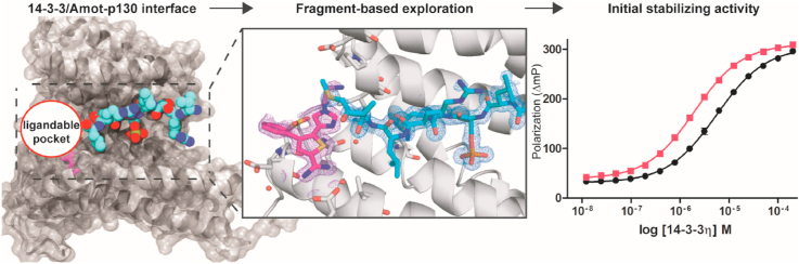
Fragment-based exploration of the 14-3-3/Amot-p130 interface
- PMID: 35036934
- PMCID: PMC8743172
- DOI: 10.1016/j.crstbi.2021.12.003
Fragment-based exploration of the 14-3-3/Amot-p130 interface
Abstract
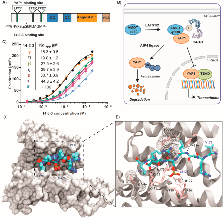
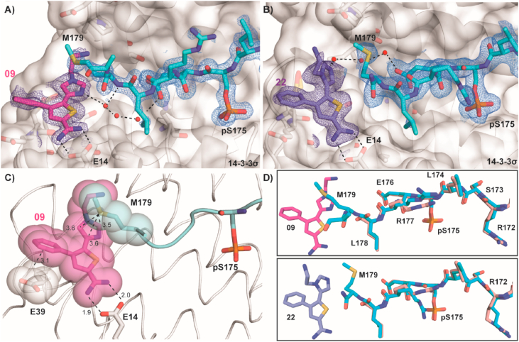
The modulation of protein-protein interactions (PPIs) has developed into a well-established field of drug discovery. Despite the advances achieved in the field, many PPIs are still deemed as 'undruggable' targets and the design of PPIs stabilizers remains a significant challenge. The application of fragment-based methods for the identification of drug leads and to evaluate the 'tractability' of the desired protein target has seen a remarkable development in recent years. In this study, we explore the molecular characteristics of the 14-3-3/Amot-p130 PPI and the conceptual possibility of targeting this interface using X-ray crystallography fragment-based screening. We report the first structural elucidation of the 14-3-3 binding motif of Amot-p130 and the characterization of the binding mode and affinities involved. We made use of fragments to probe the 'ligandability' of the 14-3-3/Amot-p130 composite binding pocket. Here we disclose initial hits with promising stabilizing activity and an early-stage selectivity toward the Amot-p130 motifs over other representatives 14-3-3 partners. Our findings highlight the potential of using fragments to characterize and explore proteins' surfaces and might provide a starting point toward the development of small molecules capable of acting as molecular glues.
Keywords: 14-3-3 /protein-protein interactions stabilizers; AIP4, Atrophin-1 interacting protein 4; Amot, Angiomotin; Amot-p130; AmotL1/2, Angiomotin-like 1/2; FBDD, Fragment-based drug discovery; FP, Fluorescence polarization; Fragment-based drug discovery; Lats 1/2, Large tumor suppressor 1/2; Ligandability; MST, Microscale thermophoresis; PPI, Protein-protein interaction; PTMs, post-translational modifications; X-ray crystallography; YAP1, Yes-associated protein 1.
© 2021 The Author(s).
Conflict of interest statement
The authors declare the following financial interests/personal relationships which may be considered as potential competing interests: LB and CO are co-founders and share-holders of Ambagon Therapeutics.
Figures


References
-
- Adams P.D., Afonine P.V., Bunkóczi G., Chen V.B., Davis I.W., Echols N., Headd J.J., Hung L.-W., Kapral G.J., Grosse-Kunstleve R.W., McCoy A.J., Moriarty N.W., Oeffner R., Read R.J., Richardson D.C., Richardson J.S., Terwilliger T.C., Zwart P.H. PHENIX: a comprehensive Python-based system for macromolecular structure solution. Acta Crystallogr. Sect. D Biol. Crystallogr. 2010;66:213–221. - PMC - PubMed
-
- Adler J.J., Johnson D.E., Heller B.L., Bringman L.R., Ranahan W.P., Conwell M.D., Sun Y., Hudmon A., Wells C.D. Serum deprivation inhibits the transcriptional Co-activator YAP and cell growth via phosphorylation of the 130-KDa isoform of Angiomotin by the LATS1/2 protein kinases. Proc. Natl. Acad. Sci. U. S. A. 2013;110:17368–17373. - PMC - PubMed
-
- Anders C., Higuchi Y., Koschinsky K., Bartel M., Schumacher B., Thiel P., Nitta H., Preisig-Müller R., Schlichthörl G., Renigunta V., Ohkanda J., Daut J., Kato N., Ottmann C. A semisynthetic fusicoccane stabilizes a protein-protein interaction and enhances the expression of K+ channels at the cell surface. Chem. Biol. 2013;20:583–593. - PubMed
-
- Andrei S.A., Sijbesma E., Hann M., Davis J., O'Mahony G., Perry M.W.D., Karawajczyk A., Eickhoff J., Brunsveld L., Doveston R.G., Milroy L.-G., Ottmann C. Stabilization of protein-protein interactions in drug discovery. Expet Opin. Drug Discov. 2017;12:925–940. - PubMed
LinkOut - more resources
Full Text Sources
Research Materials
Miscellaneous
