. 2022 Feb 22;50(3):1484-1500.
doi: 10.1093/nar/gkab1303.

Characterization of the SARS-CoV-2 ExoN (nsp14ExoN-nsp10) complex: implications for its role in viral genome stability and inhibitor identification

Affiliations

Characterization of the SARS-CoV-2 ExoN (nsp14ExoN-nsp10) complex: implications for its role in viral genome stability and inhibitor identification

Hannah T Baddock et al. Nucleic Acids Res. .

Abstract

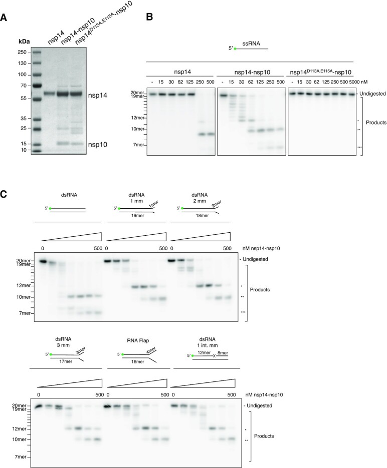
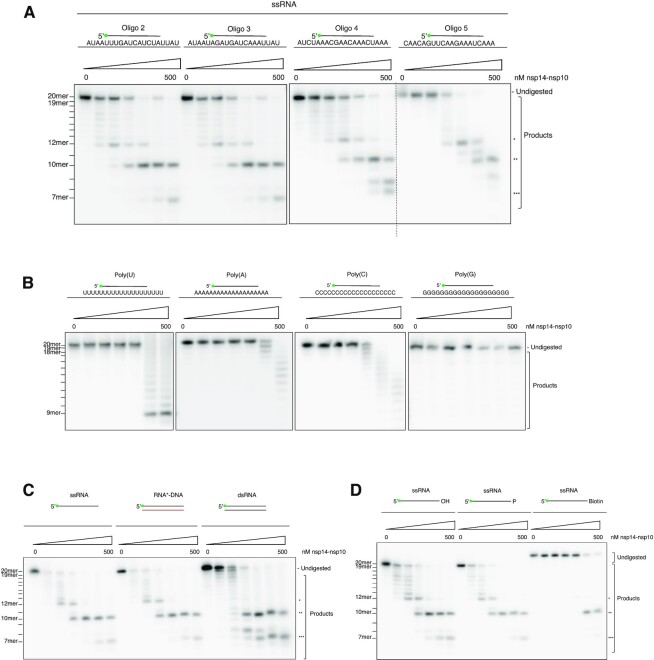
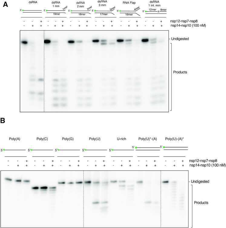
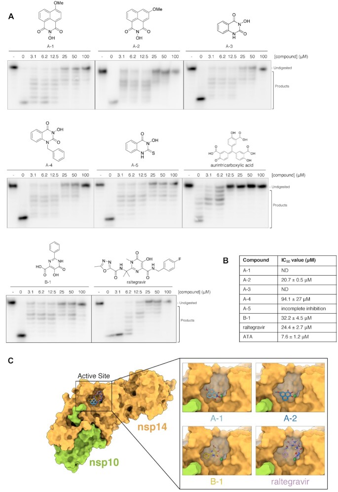
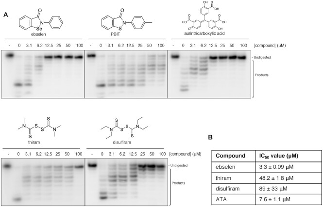
The SARS-CoV-2 coronavirus is the causal agent of the current global pandemic. SARS-CoV-2 belongs to an order, Nidovirales, with very large RNA genomes. It is proposed that the fidelity of coronavirus (CoV) genome replication is aided by an RNA nuclease complex, comprising the non-structural proteins 14 and 10 (nsp14-nsp10), an attractive target for antiviral inhibition. Our results validate reports that the SARS-CoV-2 nsp14-nsp10 complex has RNase activity. Detailed functional characterization reveals nsp14-nsp10 is a versatile nuclease capable of digesting a wide variety of RNA structures, including those with a blocked 3'-terminus. Consistent with a role in maintaining viral genome integrity during replication, we find that nsp14-nsp10 activity is enhanced by the viral RNA-dependent RNA polymerase complex (RdRp) consisting of nsp12-nsp7-nsp8 (nsp12-7-8) and demonstrate that this stimulation is mediated by nsp8. We propose that the role of nsp14-nsp10 in maintaining replication fidelity goes beyond classical proofreading by purging the nascent replicating RNA strand of a range of potentially replication-terminating aberrations. Using our developed assays, we identify drug and drug-like molecules that inhibit nsp14-nsp10, including the known SARS-CoV-2 major protease (Mpro) inhibitor ebselen and the HIV integrase inhibitor raltegravir, revealing the potential for multifunctional inhibitors in COVID-19 treatment.


Figures

Figure 1.
Figure 1.
SARS-CoV-2 nsp14–nsp10 is a versatile RNA proofreading nuclease.(A) SDS-PAGE of purified nsp14 alone and nsp14–nsp10 and an inactive control complex bearing substitutions at residues D113 and E115 (nsp14D113A,E115A–nsp10) showing the purity of the purified proteins. Predicted molecular weights are nsp14: 60 034 Da (wild-type nsp14–nsp10), 59 931 Da (nsp14D113A,E115A–nsp10) and nsp10: 14 790 Da. (B) Nsp14–nsp10 is an RNase able to digest a 20-mer ssRNA oligo (Oligo 2 in Supplementary Table 1A) in a single-nucleotide fashion from the 3'-end terminating at the eighth ribonucleotide from the 3'-end (labelled *) and further incising closer to the 5'-end to generate 10-mer and 7-mer products (labelled ** and *** respectively). Nsp14 alone is able to generate the 12-mer and 10-mer products at significantly higher protein concentrations. The predicted inactive nsp14D113A,E115A–nsp10 complex exhibits no discernible activity, even at ten-fold higher concentrations compared with the wild-type complex. (C) Nsp14–nsp10 is an exo- and endo-nuclease acting on a variety of RNA substrates, including RNA substrates with mismatched termini and flaps with no preference for mismatched ribonucleotides. Quantification in Suppl. Figure 2B. mm: mismatch. int. mm: internal mismatch. Increasing concentrations of protein (as indicated) were incubated with substrate (37°C, 45 min). Reactions were analysed by 20% denaturing PAGE. Size of products was determined as shown in Supplementary Figure 1E. Main products are labelled *, ** and *** corresponding to 12-mer, 10-mer and 7-mer respectively. All oligonuleotides used are given in Supplementary Table 1A and B.
Figure 2.
Figure 2.
Nsp14–nsp10 exhibits both 3'-exonuclease activity and a newly-described endonucleolytic activity that extends beyond the classical role of a proofreading nuclease.(A) The nuclease activity of nsp14–nsp10 is not sequence specific on a mixed-sequence substrate. The complex shows indistinguishable digestion patterns on four 20-mer ssRNA substrates of different sequences and containing all four ribonucleotides. Oligonucleotides 2, 3, 4 and 5 were used respectively (see Supplementary Table 1A). (B) When presented with 20-mer Poly(U), Poly(A), Poly(C) and Poly(G) ssRNA (oligonucleotides 11, 12, 13 and 14 respectively, see Supplementary Table 1A), nsp14–nsp10 shows reduced and qualitatively altered activity, with a single nucleotide step-wise digestion from the 3'-end curtailing at the 9th–11th nucleotide from the 3'-end. (C) Nsp14–nsp10 processes ssRNA, the RNA strand of an RNA:DNA hybrid, and dsRNA with no preference for double-stranded substrates. For all structures, the labelled oligo is oligo 2 (see Supplementary Table 1A and B). (D) Nsp14–nsp10 has RNA exo- and endo- nuclease activities. With a substrate containing a 3'-biotin group, the characteristic exonucleolytic laddering of the substrate is lost and only endonucleolytic cleavage at positions furthest from the 3'-end is observed; substrates with a 3'-hydroxyl or phosphate exhibit nearly identical product formation profiles. Oligonucleotides 2, 15 and 16 were used respectively (see Supplementary Table 1A). Increasing concentrations of protein (as indicated) were incubated with substrate (37°C, 45 min); reactions were subsequently analysed by 20% denaturing PAGE. The size of products was determined as shown in Supplementary Figure 1E. Main products are labelled *, ** and *** corresponding to 12-mer, 10-mer and 7-mer respectively. All oligonucleotides used are indicated in Supplementary Table 1A and B.
Figure 3.
Figure 3.
The SARS-CoV-2 polymerase nsp12-nsp7-nsp8 complex enhances nsp14–nsp10 nuclease activity on a variety of RNA substrates.(A) In the presence of the nsp12–7–8 polymerase complex, nsp14–nsp10 shows enhanced activity on a variety of RNA substrates, including RNA substrates with mismatched termini and flaps with no observed differential effects. mm: mismatch int mm: internal mismatch. B. When presented with 20-mer Poly(A), Poly(C), Poly(G), Poly(U) and U-rich ssRNA (oligonucleotides 12, 13, 14, 11 and 25 respectively, see Supplementary Table 1A) as well as double-stranded Poly(U)*-(A) and Poly(U)-(A)*, nsp14–nsp10 shows a more profound stimulation on repetitive sequences when nsp12–7–8 is present. Enhancement of nsp14–nsp10 nuclease activity by nsp12–7–8 is most pronounced on uracil-containing substrates. * Indicates labelled strand. The SARS-CoV-2 nsp12–7–8 polymerase complex (at a 1:3:3 molar ratio) was incubated with 10 nM substrate (37 °C, 5 min) prior to addition of 100 nM of nsp14–nsp10 at a 1:2 molar ratio of nsp14–nsp10 complex to nsp12–7–8 complex. Reactions were incubated at 37 °C for 45 min, then analysed by 20% denaturing PAGE. The size of products was determined as shown in Supplementary Figure 1E. Main products are labelled *, ** and *** corresponding to 12-mer, 10-mer and 7-mer respectively. Oligonucleotides used are given in Supplementary Table 1A and B.
Figure 4.
Figure 4.
The SARS-CoV-2 polymerase nsp12–nsp7–nsp8 complex enhances nsp14–nsp10 nuclease activity on a variety of RNA substrates.(A) The SARS-CoV-2 primase, nsp8 enhances nsp14–nsp10 activity. The addition of each individual polymerase subunit, i.e.nsp12, nsp7, and/or nsp8, as well as different combinations reveal nsp8 as the major enhancer of nsp14–nsp10 activity. Mutant nsp12D126A and nsp12D760A-D761A show no substantial stimulation of activity of nsp14–nsp10 compared to nsp8 suggesting stimulation of nuclease activity is uncoupled from polymerase activity. (B) The presence of nsp8 stimulates the exonuclease activity of SARS-CoV-2 nsp14–nsp10. Increasing concentrations of nsp14–nsp10 were incubated with ss- or dsRNA (Oligo 2 and 3, see Supplementary Table 1A) and a fixed concentration of 300 nM nsp8 (37°C, 45 min). Reactions were analysed by 20% denaturing PAGE. (C) The presence of nsp8 stimulates the endonuclease activity of nsp14–nsp10. Increasing concentrations of nsp14–nsp10 were incubated with ss- or dsRNA with a blocked (3′-biotin) terminus (Oligo 2 and 16 see Supplementary Table 1A) and a fixed concentration of 300 nM nsp8 at (37°C, 45 min). Reactions were analysed by 20% denaturing PAGE. The SARS-CoV-2 nsp12–7–8 polymerase complex (at a 1:3:3 molar ratio) was incubated with 10 nM substrate (37°C, 5 min), prior to addition of 100 nM of nsp14–nsp10 at a 1:2 molar ratio of nsp14–nsp10 complex to nsp12–7–8 complex. Reactions were incubated at 37°C for 45 min and subsequently analysed by 20% denaturing PAGE. The size of products was determined as shown in Supplementary Figure 1E. Main products are labelled *, ** and *** corresponding to 12-mer, 10-mer and 7-mer respectively. Oligonucleotides used are given in Supplementary Table 1A and B.
Figure 5.
Figure 5.
The exonuclease activity of nsp14–nsp10 is inhibited by N-hydroxyimide and hydroxypyrimidinone based compounds. (A) Increasing concentrations (as indicated, in μM) of compounds incubated with 100 nM nsp14–nsp10 (room temperature, 10 min), before initiating nuclease reaction by addition of ssRNA (37 °C, 45 min). Products were analysed by 20% denaturing PAGE. A decrease in the generation of nucleolytic reaction products and an increase in undigested substrate indicates inhibition of nuclease activity at increasing inhibitor concentrations. - indicates no enzyme. Compounds A-1–A-4 are based on a N-hydroxyimide scaffold, B-1 is a hydroxypyrimidinone. (B) IC50 values determined by quantification of gel digestion products (100 nM nsp14–nsp10); dose-response curves were determined by nonlinear regression. The mean ± s.e.m. were calculated from ≥3 biological repeats. (C) Docking of nsp14–nsp10 using Autodock. Nsp14–nsp10 was docked with compounds within grid boxes encompassing a surface focussed on the active site surface then the highest-affinity docking pose of A-1 and A-2 overlaid on the surface of SARS-CoV nsp14–nsp10; Mg2+ is in dark green and the highest-affinity docking poses of B1 and raltegravir overlaid on the surface of SARS-CoV nsp14–nsp10. Nsp14 is in yellow-orange, nsp10 is in light green, Mg2+ is in dark green. The docked poses of the compounds on the surface of the whole nsp14–nsp10 complex are shown on the left-hand side, with a detailed view at the active site on the right-hand side inset.
Figure 6.
Figure 6.
The exonuclease activity of nsp14–nsp10 is inhibited by the presence of drugs and drug-like compounds. (A) Increasing concentrations (as indicated, in μM) of drug and drug-like compounds were incubated with 100 nM nsp14–nsp10 (room temperature, 10 min), before initiation of nuclease reaction by the addition of ssRNA (37°C, 45 min). Products were analysed by 20% denaturing PAGE. A decrease in the generation of nucleolytic reaction products and a concomitant increase in undigested substrate indicates inhibition of nuclease activity. – indicates no enzyme. Gels are representative of at least three biological repeats. (B) IC50 values as calculated by quantification of gel digestion products (100 nM nsp14–nsp10) and dose-response curves were determined by nonlinear regression. The mean ± s.e.m. were calculated from ≥3 biological repeats. Precise IC50 values should be regarded as preliminary due to the nature of the assay.

References

    1. Fung T.S., Liu D.X.. Human coronavirus: host-pathogen interaction. Annu Rev Microbiol. 2019; 73:529–557. - PubMed
    1. Sevajol M., Subissi L., Decroly E., Canard B., Imbert I.. Insights into RNA synthesis, capping, and proofreading mechanisms of SARS-coronavirus. Virus Res. 2014; 194:90–99. - PMC - PubMed
    1. Bouvet M., Imbert I., Subissi L., Gluais L., Canard B., Decroly E.. RNA 3'-end mismatch excision by the severe acute respiratory syndrome coronavirus nonstructural protein nsp10/nsp14 exoribonuclease complex. Proc. Natl. Acad. Sci. U.S.A. 2012; 109:9372–9377. - PMC - PubMed
    1. Eckerle L.D., Lu X., Sperry S.M., Choi L., Denison M.R.. High fidelity of murine hepatitis virus replication is decreased in nsp14 exoribonuclease mutants. J. Virol. 2007; 81:12135–12144. - PMC - PubMed
    1. Smith E.C., Blanc H., Surdel M.C., Vignuzzi M., Denison M.R.. Coronaviruses lacking exoribonuclease activity are susceptible to lethal mutagenesis: evidence for proofreading and potential therapeutics. PLoS Pathog. 2013; 9:e1003565. - PMC - PubMed

Publication types

MeSH terms