. 2022 Jan 26;144(3):1332-1341.
doi: 10.1021/jacs.1c11434. Epub 2022 Jan 17.

Faster Crystallization during Coral Skeleton Formation Correlates with Resilience to Ocean Acidification

Affiliations

Faster Crystallization during Coral Skeleton Formation Correlates with Resilience to Ocean Acidification

Connor A Schmidt et al. J Am Chem Soc. .

Abstract

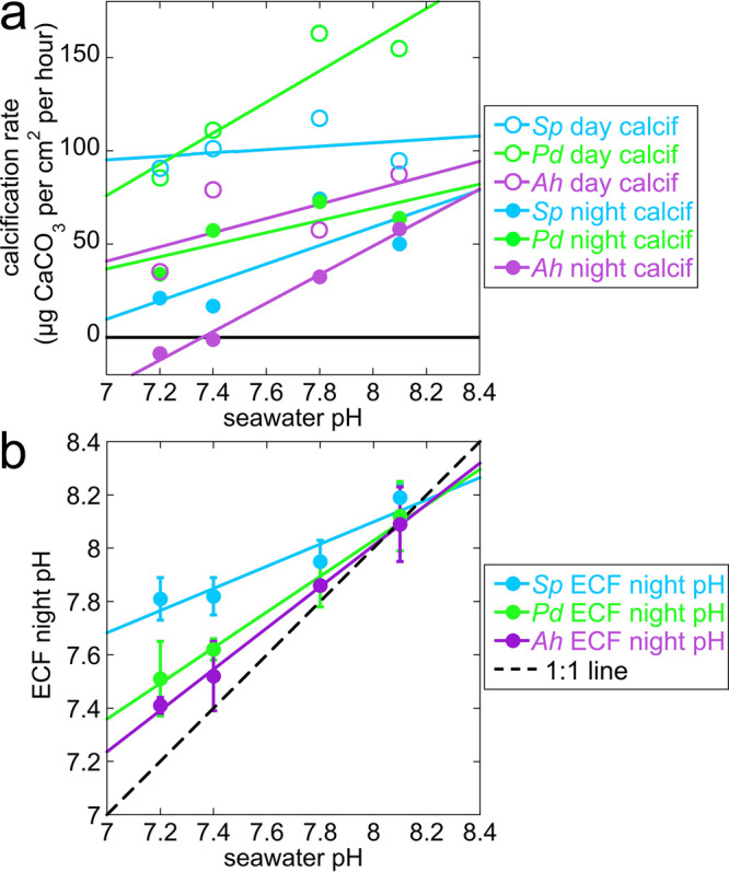
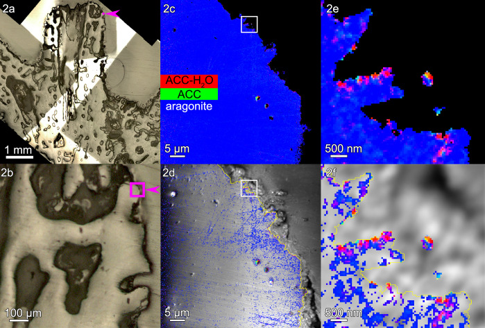
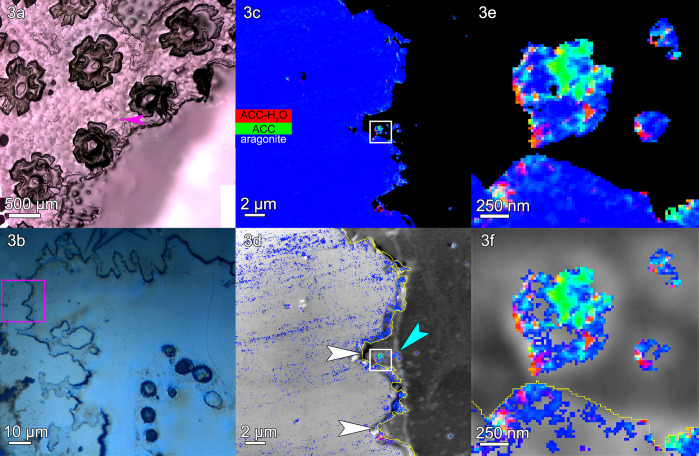
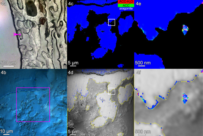
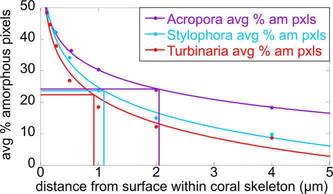
The mature skeletons of hard corals, termed stony or scleractinian corals, are made of aragonite (CaCO3). During their formation, particles attaching to the skeleton's growing surface are calcium carbonate, transiently amorphous. Here we show that amorphous particles are observed frequently and reproducibly just outside the skeleton, where a calicoblastic cell layer envelops and deposits the forming skeleton. The observation of particles in these locations, therefore, is consistent with nucleation and growth of particles in intracellular vesicles. The observed extraskeletal particles range in size between 0.2 and 1.0 μm and contain more of the amorphous precursor phases than the skeleton surface or bulk, where they gradually crystallize to aragonite. This observation was repeated in three diverse genera of corals, Acropora sp., Stylophora pistillata─differently sensitive to ocean acidification (OA)─and Turbinaria peltata, demonstrating that intracellular particles are a major source of material during the additive manufacturing of coral skeletons. Thus, particles are formed away from seawater, in a presumed intracellular calcifying fluid (ICF) in closed vesicles and not, as previously assumed, in the extracellular calcifying fluid (ECF), which, unlike ICF, is partly open to seawater. After particle attachment, the growing skeleton surface remains exposed to ECF, and, remarkably, its crystallization rate varies significantly across genera. The skeleton surface layers containing amorphous pixels vary in thickness across genera: ∼2.1 μm in Acropora, 1.1 μm in Stylophora, and 0.9 μm in Turbinaria. Thus, the slow-crystallizing Acropora skeleton surface remains amorphous and soluble longer, including overnight, when the pH in the ECF drops. Increased skeleton surface solubility is consistent with Acropora's vulnerability to OA, whereas the Stylophora skeleton surface layer crystallizes faster, consistent with Stylophora's resilience to OA. Turbinaria, whose response to OA has not yet been tested, is expected to be even more resilient than Stylophora, based on the present data.


Conflict of interest statement

The authors declare no competing financial interest.

Figures

Figure 1
Figure 1
(a) Calcification rate and (b) night-time pH measured in extracellular calcifying fluid (ECF), between calicoblastic epithelium and forming skeleton surface, as seawater pH decreases in simulated ocean acidification (OA, pH 8.1 → 7.2) experiments in three coral genera and species: Stylophora pistillata (Sp, light blue), Pocillopora damicornis (Pd, green), and Acropora hyacinthus (Ah, purple). These are selected, replotted data from Venn et al., 2019. (a) During the day (open circles) the calcification rate is constant for Stylophora, but it decreases with OA for Pocillopora and Acropora. At night, calcification decreases with OA for all genera, but especially for Acropora, which goes below zero (black solid line); thus, the skeleton formed during the day dissolves at night. (b) The pH values in the ECF during the day, when photosynthesis is active, decrease to 7.8 identically for all three genera; thus, they are omitted here. Only night-time pH values in the ECF are shown, as they vary dramatically across the three genera. The solid lines are linear fits of the data; the 1:1 line (black dashed line) is where pHECF = pHseawater. Clearly, as the seawater pH decreases, the ECF night-time pH decreases, but at slower rates for all genera compared to seawater. Stylophora ECF pH is the slowest, Pocillopora intermediate, and Acropora the fastest, that is, closest to the seawater pH decrease with OA.
Figure 2
Figure 2
Acropora sp.: (a, b) polarized light microscopy (PLM) images and (c) a component map with a black mask where the Ca signal is undetectable and pixels colored according to the mineral phase spectroscopically observed. In this and all other component maps in this work, red pixels are ACC-H2O, green pixels are ACC, and blue pixels are aragonite. The component spectra used to obtain all component maps are shown in Figure S1. (d) Average PEEM image overlaid with the component map, with both black mask and pure blue aragonite removed. (e, f) One region of interest, boxed in panels c and d, magnified here to show the precise locations of both intra- and extraskeletal amorphous pixels. Extraskeletal particles are no farther than 2 μm outside the skeleton’s surface (yellow line in panels d and f), are mostly amorphous, and are assumed to be inside the calicoblastic epithelium. Intraskeletal amorphous pixels in panel d extend several micrometers inside the yellow line. See Figure S2 for more images of this area.
Figure 3
Figure 3
Stylophora pistillata: (a) PLM image, (b) DIC image, (c) a component map, and (d) an average PEEM image overlaid with the component map, as described in Figure 2. (e, f) The regions boxed in panels c and d are magnified here to show amorphous particles where calicoblastic cells are expected. This region shows several scars left by desmocytes, the cells that bind the calicoblastic epithelium to the skeleton and form 3-μm-deep V-shaped scars, indicated by white arrows in panel d. Distinct particles are visible just outside the skeleton surface (e.g., cyan arrowhead in panel d, and three particles in panel f). These extraskeletal particles have both a greater percentage of amorphous pixels and a greater concentration of amorphous phases per pixel, compared to the skeleton surface or bulk (Tables S2 and S3). Extraskeletal particles are mostly amorphous within the calicoblastic epithelium, <2 μm outside the yellow line in panels d and f. Intraskeletal amorphous pixels in panel d extend ∼1 μm inside the yellow line. See Figure S3 for more images of this area.
Figure 4
Figure 4
Turbinaria peltata: (a) PLM image, (b) Differential Interference Contrast (DIC) image, (c) component map, and (d) average PEEM image overlaid with the component map, as described in Figure 2. (e, f) The regions boxed in panels c and d are magnified here to show amorphous particles where calicoblastic cells are expected. Amorphous pixels also appear along the edge of the skeleton. Extraskeletal particles are mostly amorphous within the calicoblastic epithelium, <2 μm outside the yellow line in panels d and f. Intraskeletal amorphous pixels in panel f extend <1 μm inside the yellow line. See Figure S4 for more images of this area.
Figure 5
Figure 5
Amorphous, soluble thickness decays with distance from the skeleton surface. Comparison of the three genera analyzed here, for percentage of amorphous pixels (either ACC-H2O or ACC) as a function of distance from the surface, indicated by a yellow line in Figures 2–4. Each percentage is averaged over the five areas analyzed per genus (Table S4), the averages are displayed as circles, and the solid lines are logarithmic decays (among others tested, a logarithmic decay provided better fits, with R > 0.98). The half-lengths (vertical lines) are the distances at which the amorphous pixels have decayed to 50% of the surface value for that genus (horizontal lines). The half-lengths are 2.1 μm for Acropora, 1.1 μm for Stylophora, and 0.9 μm for Turbinaria.

References

    1. Eyre B. D.; Cyronak T.; Drupp P.; De Carlo E. H.; Sachs J. P.; Andersson A. J. Coral reefs will transition to net dissolving before end of century. Science 2018, 359 (6378), 908–911. 10.1126/science.aao1118. - DOI - PubMed
    1. Venn A.; Tambutté E.; Caminiti-Segonds N.; Techer N.; Allemand D.; Tambutté S. Effects of light and darkness on pH regulation in three coral species exposed to seawater acidification. Sci. Rep 2019, 9 (1), 2201.10.1038/s41598-018-38168-0. - DOI - PMC - PubMed
    1. Mass T.; Giuffre A. J.; Sun C.-Y.; Stifler C. A.; Frazier M. J.; Neder M.; Tamura N.; Stan C. V.; Marcus M. A.; Gilbert P. U. P. A. Amorphous calcium carbonate particles form coral skeletons. Proc. Natl. Acad. Sci. 2017, 114 (37), E7670–E7678. 10.1073/pnas.1707890114. - DOI - PMC - PubMed
    1. Sun C.-Y.; Stifler C. A.; Chopdekar R. V.; Schmidt C. A.; Parida G.; Schoeppler V.; Fordyce B. I.; Brau J. H.; Mass T.; Tambutté S.; Gilbert P. U. P. A. From particle attachment to space-filling coral skeletons. Proc. Natl. Acad. Sci. 2020, 117 (48), 30159–30170. 10.1073/pnas.2012025117. - DOI - PMC - PubMed
    1. Tambutté E.; Allemand D.; Zoccola D.; Meibom A.; Lotto S.; Caminiti N.; Tambutté S. Observations of the tissue-skeleton interface in the scleractinian coral Stylophora pistillata. Coral Reefs 2007, 26 (3), 517–529. 10.1007/s00338-007-0263-5. - DOI

Substances