. 2022 May;31(5):807-813.
doi: 10.1111/exd.14530. Epub 2022 Feb 4.

Transient receptor potential vanilloid 3 expression is increased in non-lesional skin of atopic dermatitis patients

Affiliations

Transient receptor potential vanilloid 3 expression is increased in non-lesional skin of atopic dermatitis patients

Nikolett Vasas et al. Exp Dermatol. 2022 May.

Abstract

TRPV3 (transient receptor potential vanilloid 3) is a pro-inflammatory ion channel mostly expressed by keratinocytes of the human skin. Previous studies have shown that the expression of TRPV3 is markedly upregulated in the lesional epidermis of atopic dermatitis (AD) patients suggesting a potential pathogenetic role of the ion channel in the disease. In the current study, we aimed at defining the molecular and functional expression of TRPV3 in non-lesional skin of AD patients as previous studies implicated that healthy-appearing skin in AD is markedly distinct from normal skin with respect to terminal differentiation and certain immune function abnormalities. By using multiple, complementary immunolabelling and RT-qPCR technologies on full-thickness and epidermal shave biopsy samples from AD patients (lesional, non-lesional) and healthy volunteers, we provide the first evidence that the expression of TRPV3 is markedly upregulated in non-lesional human AD epidermis, similar to lesional AD samples. Of further importance, by using the patch-clamp method on cultured healthy and non-lesional AD keratinocytes, we also show that this upregulation is functional as determined by the significantly augmented TRPV3-specific ion current (induced by agonists) on cultured non-lesional AD keratinocytes when compared to healthy ones.

Keywords: TRPV3; atopic dermatitis; inflammatory skin diseases; keratinocytes.


Conflict of interest statement

TB provides consultancy services to Phytecs, Inc. (Los Angeles, CA, USA) and Monasterium Laboratory (Münster, Germany) who had no role in conceiving the study, designing the experiments, writing the manuscript or in the decision to publish it. The other authors state no conflict of interest.

Figures

FIGURE 1
FIGURE 1
Expression of TRPV3 is significantly increased in AD patients. (A) TRPV3‐specific immunoreactivity (IR), as shown by immunohistochemistry on representative healthy, non‐lesional and lesional atopic dermatitis (AD) skin sections. Magnification 200x (top row) and 400x (bottom row). Scale bars = 50 μm. (B) Semiquantitative analysis of TRPV3 immunoreactivity on 4 histological sections per group. Results are expressed as mean ± SD. (C, E) TRPV3 mRNA expression in healthy human (Healthy) as well as non‐lesional and lesional AD (Non‐Lesional and Lesional) skin tissue, and cell lysates of keratinocytes isolated from healthy human (NHEK: normal human epidermal keratinocyte) and non‐lesional and lesional AD (AD‐NHEK: Non‐Lesional human epidermal keratinocyte from atopic dermatitis patient and AD‐HEK: Lesional human epidermal keratinocyte from atopic dermatitis patient, 70%: samples harvested at 70% confluence, PC2: samples harvested on day 2 after 100% confluence, respectively) samples, as assessed by quantitative real‐time PCR. Results are expressed as mean ± SD, two additional experiments showed similar results (Figure S2 and S4). (D, F) TRPV3 protein level as determined by Western blot. Optical density was normalized to β‐actin. Results are expressed as mean ± SD of 3 independent determinations. *p < 0.05, ***p < 0.001 compared to Healthy groups, #p < 0.05, ###p < 0.001 compared with the Non‐Lesional group
FIGURE 2
FIGURE 2
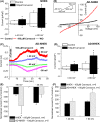
Function of TRPV3 is significantly increased in AD patients. (A) Representative I‐V curves recorded in AD‐NHEK cells in whole‐cell patch‐clamp configuration, in control (black) and after 100 μM carvacrol (red) and washout (grey). Inset shows the patch‐clamp ramp protocol. (B) Statistical analysis of current densities normalized to cell membrane capacitance measured at −40 mV (left side downward), at +40 mV (left side upward), at −90 mV (right side downward) and at +90 mV (right side upward) in control (empty columns) and after 100 μM carvacrol (filled columns) in NHEK cells. Values are mean ± SEM, n = 14. (C) Representative experiment shows the typical time course of 100 μM carvacrol‐induced current, measured at −90 mV (magenta), at −40 mV (green), at +40 mV (blue) and at +90 mV (red). Dashed lines show changes of the experimental solutions. (D) Statistical analysis of current densities normalized to cell membrane capacitance measured at −40 mV (left side downward), at +40 mV (left side upward), at −90 mV (right side downward) and at +90 mV (right side upward) in control (empty columns) and after 100 μM carvacrol (filled columns) in AD‐NHEK cells. Values are mean ± SEM, n = 4. (E, F) Mean values show changes in currents after 100 μM carvacrol treatment. Filled columns show NHEK, while dashed columns show AD‐NHEK cells. Currents are displayed as the percentage of the control (no drug), where (E) shows currents at −90 mV and at −40 mV, while (F) shows currents at +40 mV and at +90 mV. Values are mean ± SEM, n = 14 and n = 4 for NHEK and AD‐NHEK, respectively. *p < 0.05 compared to control or NHEK groups

References

    1. Khalil M, Alliger K, Weidinger C, et al. Functional role of transient receptor potential channels in immune cells and epithelia. Front Immunol. 2018;9:174. doi:10.3389/fimmu.2018.00174 - DOI - PMC - PubMed
    1. Tóth BI, Oláh A, Szöllősi AG, Bíró T. TRP channels in the skin. Br J Pharmacol. 2014;171(10):2568‐2581. doi:10.1111/bph.12569 - DOI - PMC - PubMed
    1. Peier AM, Reeve AJ, Andersson DA, et al. A heat‐sensitive TRP channel expressed in keratinocytes. Science. 2002;296(5575):2046‐2049. doi:10.1126/science.1073140 - DOI - PubMed
    1. Asakawa M, Yoshioka T, Matsutani T, et al. Association of a mutation in TRPV3 with defective hair growth in rodents. J Invest Dermatol. 2006;126(12):2664‐2672. doi:10.1038/sj.jid.5700468 - DOI - PubMed
    1. Yoshioka T, Imura K, Asakawa M, et al. Impact of the Gly573Ser substitution in TRPV3 on the development of allergic and pruritic dermatitis in mice. J Invest Dermatol. 2009;129(3):714‐722. doi:10.1038/jid.2008.245 - DOI - PubMed

Publication types