Review
. 2022 Jul;162(1):156-178.
doi: 10.1016/j.chest.2021.12.659. Epub 2022 Jan 14.

Algorithmic Approach to the Diagnosis of Organizing Pneumonia: A Correlation of Clinical, Radiologic, and Pathologic Features

Affiliations
Review

Algorithmic Approach to the Diagnosis of Organizing Pneumonia: A Correlation of Clinical, Radiologic, and Pathologic Features

Sujith V Cherian et al. Chest. 2022 Jul.

Abstract

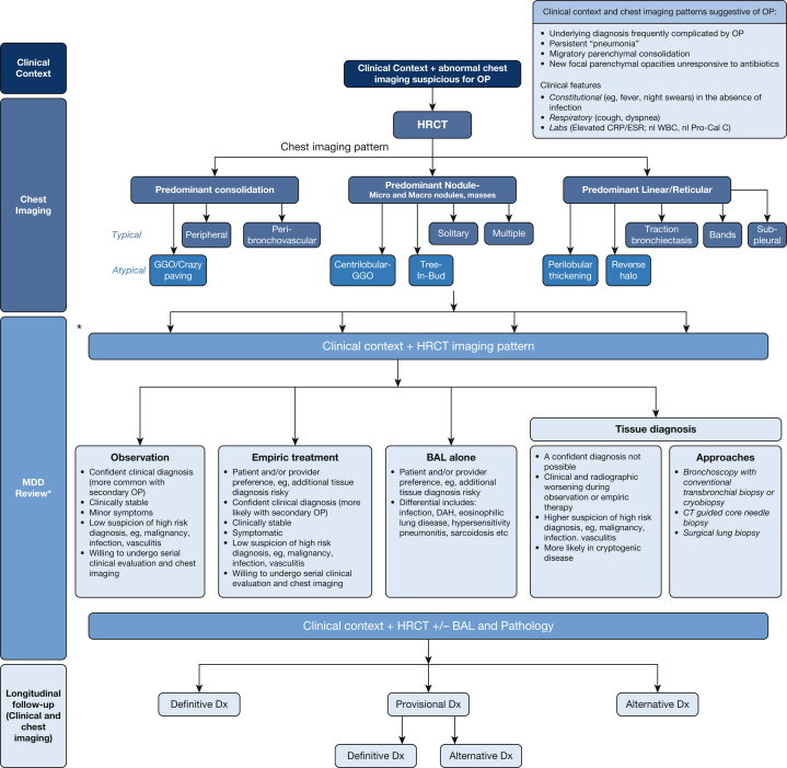
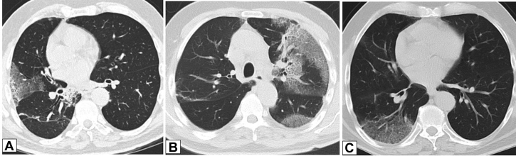
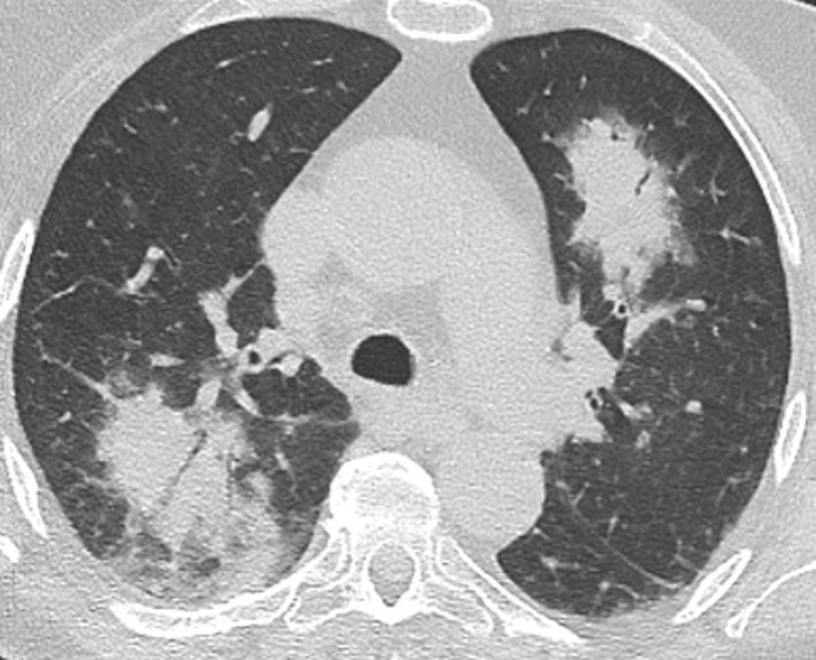
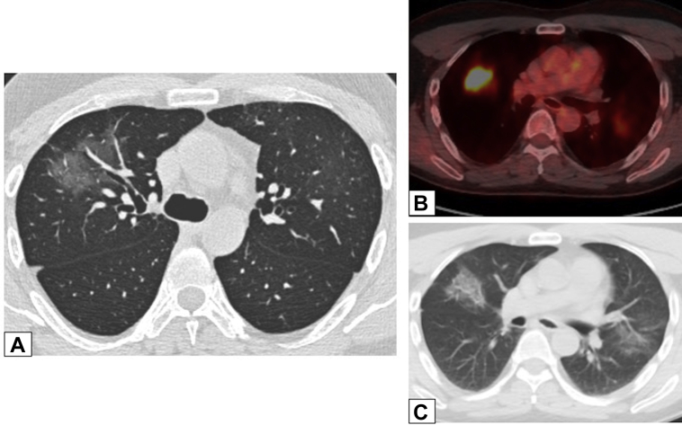
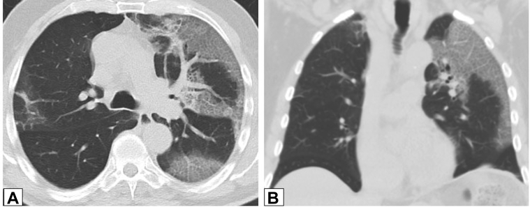
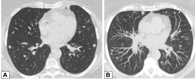
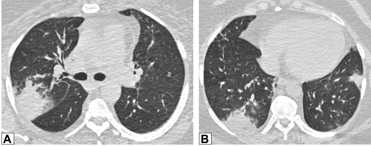
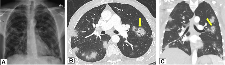
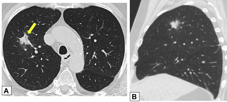
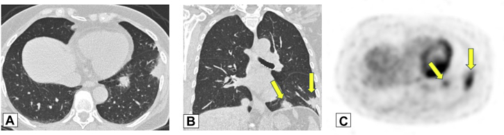
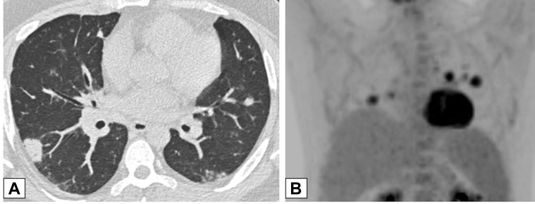
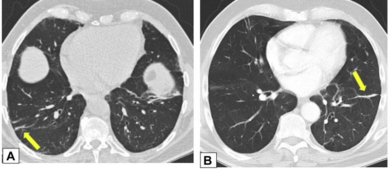
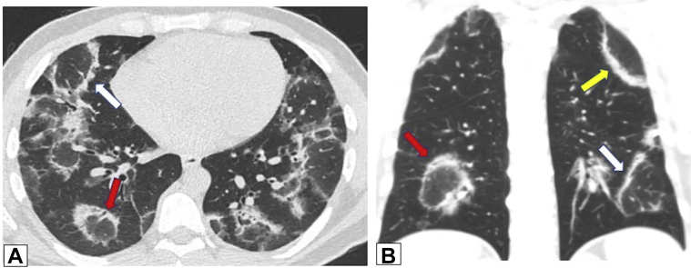
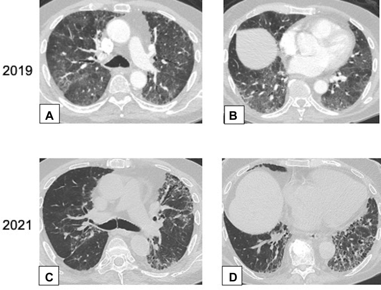
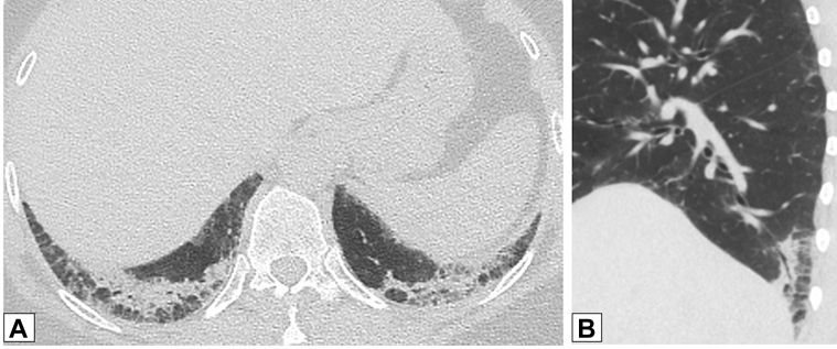
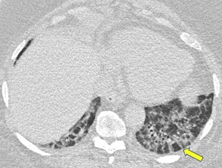
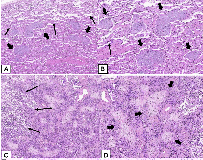
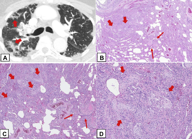
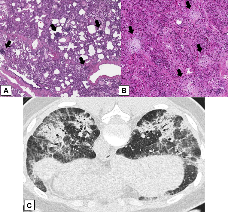
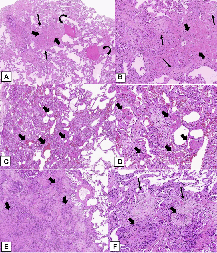
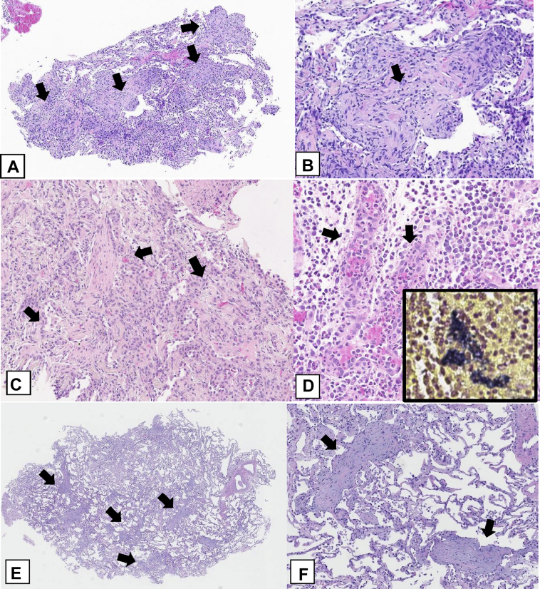
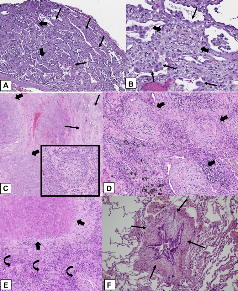
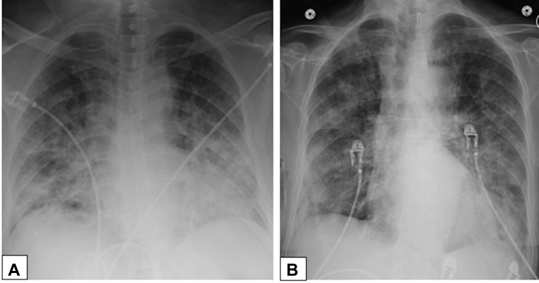
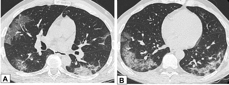
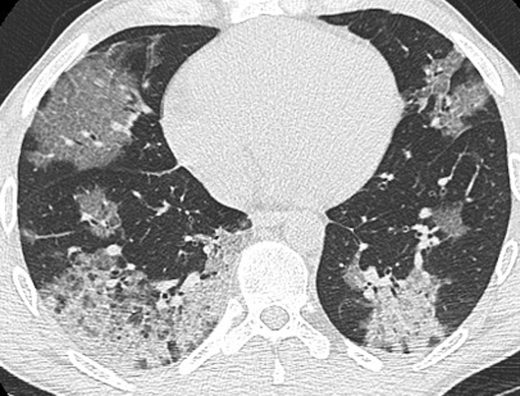
Organizing pneumonia (OP), characterized histopathologically by patchy filling of alveoli and bronchioles by loose plugs of connective tissue, may be seen in a variety of conditions. These include but are not limited to after an infection, drug reactions, radiation therapy, and collagen vascular diseases. When a specific cause is responsible for this entity, it is referred to as "secondary OP." When an extensive search fails to reveal a cause, it is referred to as "cryptogenic OP" (previously called "bronchiolitis obliterans with OP"), which is a clinical, radiologic, and pathologic entity classified as an interstitial lung disease. The clinical presentation of OP often mimics that of other disorders, such as infection and cancer, which can result in a delay in diagnosis and inappropriate management of the underlying disease. The radiographic presentation of OP is polymorphous but often has subpleural consolidations with air bronchograms or solitary or multiple nodules, which can wax and wane. Diagnosis of OP sometimes requires histopathologic confirmation and exclusion of other possible causes. Treatment usually requires a prolonged steroid course, and disease relapse is common. The aim of this article is to summarize the clinical, radiographic, and histologic presentations of this disease and to provide a practical diagnostic algorithmic approach incorporating clinical history and characteristic imaging patterns.

Keywords: acute fibrinous and organizing pneumonia; cryptogenic organizing pneumonia; focal organizing pneumonia; organizing pneumonia; secondary organizing pneumonia.


Figures

Figure 1
Figure 1
Algorithmic approach to organizing pneumonia. ∗ A formal MDD may not be required in all cases, especially if the combination of clinical context and radiographic pattern is sufficiently convincing of the OP diagnosis. In such cases, a discussion between the physician and the radiologist is strongly encouraged. CRP = C-reactive protein; DAH = diffuse alveolar hemorrhage; Dx = diagnosis; ESR = erythrocyte sedimentation rate; GGO = ground-glass opacification; HRCT = high-resolution CT; MDD = multidisciplinary discussion; nl Pro-Cal C = normal procalcitonin; nl WBC = normal WBC; OP = organizing pneumonia.
Figure 2
Figure 2
A, B, Peripheral consolidation. Axial unenhanced CT scan images obtained in two patients. A, This image shows peripheral consolidation in the right upper lobe in a woman previously treated with radiation for breast cancer and biopsy-proved organizing pneumonia (OP). B, This image shows peripheral consolidation in the left lower lobe and lingula and biopsy-proved OP secondary to chemotherapy for treatment of lymphoma.
Figure 3
Figure 3
A, B, Peribronchovascular consolidation. Axial contrast-enhanced CT scan images through the middle (A) and lower (B) lung zones show multifocal, bilateral foci of peribronchovascular consolidation in a patient with biopsy-proved organizing pneumonia.
Figure 4
Figure 4
A-C, Migratory manifestation of organizing pneumonia (OP). A, Posteroanterior chest radiograph in a 71-year-old man presenting with dyspnea on exertion in July 2011 shows peripheral right upper lobe (RUL) consolidation. B, Repeat study from June 2012 shows that the RUL abnormality had resolved and that there is now extensive consolidation in the left lung. C, In June 2013, the left lung is now clear, but there is now new right lower lobe consolidation. These findings are typical of a migratory manifestation of OP.
Figure 5
Figure 5
A-C, Migratory manifestation of organizing pneumonia (OP). Axial unenhanced CT scans were obtained in a 71-year-old man with dyspnea on exertion, the same patient as in Figure 4. The CT scanning sections, which correspond with Figures 4A, 4B, and 4C, respectively, show that foci of consolidation on the chest radiographs in this case are due to migratory foci of ground-glass opacification with interlobular septal thickening and intralobular lines (crazy paving). Surgical lung biopsy results were consistent with OP.
Figure 6
Figure 6
Multifocal, peribronchovascular, mass-like consolidation. Axial unenhanced CT scan image shows multifocal, peribronchovascular, mass-like consolidation in a 70-year-old woman with persistent shortness of breath 9 months after the diagnosis of COVID-19 pneumonia. Organizing pneumonia was presumed to be the diagnosis after dramatic improvement in symptoms and resolution of consolidation after treatment with steroids.
Figure 7
Figure 7
A-C, Ground-glass opacification (GGO). A, Axial unenhanced CT scan image in a 47-year-old man with a history of papillary thyroid cancer, 4 months after treatment with iodine-131, shows right upper lobe peribronchovascular GGO. The GGO is hypermetabolic on a fluorine-18 fluorodeoxyglucose-PET/CT scan (B), with less intense activity also noted in a new smaller area of GGO in the left upper lobe in an axial unenhanced CT scan image (C). Transbronchial biopsy results were consistent with organizing pneumonia.
Figure 8
Figure 8
A, B, Crazy-paving pattern. Axial (A) and coronal (B) unenhanced CT scan images in a 71-year-old man who had secondary organizing pneumonia due to amiodarone use. There is extensive ground-glass opacification in the left lung with superimposed interlobular septal thickening and intralobular lines (crazy paving).
Figure 9
Figure 9
A, B, Small micronodules. Axial high-resolution CT scanning section (A) and maximum intensity projection image (B) in the same patient. CT scan images demonstrate multiple small, solid lung nodules bilaterally that are suggestive of possible malignancy or infection. CT scanning-guided core biopsy results were consistent with a diagnosis of organizing pneumonia.
Figure 10
Figure 10
A, B, Masses. Axial unenhanced CT scan images through the right upper lobe (A) and right lower lobe (B) in a 42-year-old patient who was morbidly obese and had dyspnea show bilateral, subpleural or peripheral mass-like areas of consolidation. The findings are nonspecific; subsequent biopsy results documented organizing pneumonia, and findings resolved after treatment with steroids.
Figure 11
Figure 11
A-C, Air bronchograms. A 38-year-old man with ulcerative colitis treated with mesalamine developed shortness of breath. A, A posterolateral chest radiograph demonstrates multiple bilateral lung nodules. Axial (B) and coronal (C) contrast-enhanced chest CT scans show the nodules to be both peripheral and peribronchovascular, some associated with dilated airways (arrows in B and C). Core biopsy results were consistent with organizing pneumonia.
Figure 12
Figure 12
A, B, Solitary nodules. Axial (A) and sagittal (B) unenhanced images from low-dose CT scanning lung cancer screening in a 73-year-old male smoker show a solitary, irregular right upper lobe nodular opacity associated with subtle dilated airways (arrow in A). Endobronchial ultrasound-guided fine-needle biopsy results were consistent with focal organizing pneumonia. The opacity resolved without treatment on a follow-up scan (not shown).
Figure 13
Figure 13
A-C, Fluorine-18 fluorodeoxyglucose (FDG)-PET scanning. Axial (A) and coronal (B) unenhanced CT scan images from a routine surveillance scan in a 70-year-old woman without symptoms with a history of breast cancer and long-term nitrofurantoin use show irregular, solid lung nodules in both lower lobes. C, FDG-PET scanning was performed because of concern for metastatic disease, and an axial FDG-PET image shows the left lower lobe (LLL) nodules (arrows in B and C) to be FDG avid. Core transthoracic needle biopsy results for the LLL nodule were consistent with organizing pneumonia.
Figure 14
Figure 14
A, B, Fluorine-18 fluorodeoxyglucose (FDG)-PET scanning. A 34-year-old woman with systemic lupus erythematosus presented with shortness of breath. A, An axial unenhanced chest CT scan through the lower lobes shows multiple variable-sized solid nodules in both lungs, including micronodular changes in the left base. B, A coronal image from FDG-PET scanning shows the nodules are hypermetabolic. Wedge biopsy results from two nodules were consistent with organizing pneumonia.
Figure 15
Figure 15
A, B, Parenchymal bands. Axial unenhanced (A) and contrast-enhanced (B) CT scans obtained 1 year apart in same patient with biopsy-proved organizing pneumonia show only partially resolving parenchymal bands (arrows in A and B) in both lower lobes.
Figure 16
Figure 16
A, B, Reverse halo or atoll sign. CT scan images obtained in a 32-year-old man with chronic persistent cough and dyspnea on exertion for 1 year and 20-pound weight loss. Axial (A) and coronal (B) images show bilateral foci of central ground-glass opacification completely surrounded by dense peripheral consolidation—the reverse halo or atoll sign (red arrows in A and B). Also present are foci of incomplete halo signs (white arrows in A and B) and perilobular thickening (yellow arrow in B). Overlap of these findings is clearly present. Lung biopsy results documented organizing pneumonia.
Figure 17
Figure 17
A-D, Interstitial fibrosis. Axial CT scan images obtained in a 73-year-old man after a 4-day onset of dyspnea. The CT scan images of the chest were obtained with lung windows at the level of the carina (A) and (C) and at the lung bases (B) and (D), respectively. Contrast-enhanced images in the upper row from 2019 (A and B) show extensive ground-glass opacification (GGO) bilaterally. Unenhanced images in the lower row from 2021 (C and D) show improvement in the extent of GGO but development of increased reticulation and traction bronchiectasis or bronchiolectasis, a pattern consistent with fibrosis. Wedge biopsy results from the right upper, middle, and lower lobes showed organizing pneumonia.
Figure 18
Figure 18
A, B, Nonspecific interstitial pneumonia (NSIP). Unenhanced CT scans were obtained in a 51-year-old woman with dermatomyositis. Axial (A) and sagittal (B) images through the posterior lung bases show consolidation in both lower lobes with subpleural sparing and perilobular opacities bilaterally. Dilated bronchi were evident on the sagittal image (B). The findings were consistent with overlap of organizing pneumonia and NSIP.
Figure 19
Figure 19
Nonspecific interstitial pneumonia (NSIP). An axial CT scan was obtained in a 67-year-old man who was morbidly obese and had progressive dyspnea. The CT scan through the lung bases shows a combination of thick-walled peripheral bronchi associated with poorly defined ground-glass attenuation and perilobular thickening, especially in the left base (arrow), which are findings suggestive of NSIP. Surgical lung biopsy findings were consistent with organizing pneumonia.
Figure 20
Figure 20
A-D, Histologic findings of an organizing pneumonia (OP) pattern (OPP). In this case of cryptogenic OP with multifocal lung involvement, a low-power histologic image (A) shows patchy areas of polypoid intraluminal plugs (thick arrows) of loose connective tissue that protrude into distal airways. The adjacent lung parenchyma is relatively normal (thin arrows). B, In this field, the polypoid plugs are seen within alveolar spaces and alveolar ducts (thick arrows), but bronchiolar involvement is minimal (thin arrows). The connective tissue is the same age, and the alveolar architecture is preserved. C, In this case of focal OP, there is a nodular lesion consisting of the OPP as seen in (A) and (B) surrounded by relatively normal lung (thin arrows). D, The lesion consists of polypoid plugs of loose connective tissue within distal airspaces (thick arrows).
Figure 21
Figure 21
A-D, Perilobular organizing pneumonia (OP) pattern (OPP). A, Axial CT scan image obtained at the level of the upper lobes shows perilobular opacities in the periphery of the right lung (arrows). B, In this surgical lung biopsy specimen, the lesions of the OPP (thick arrows) are also seen histologically to be situated at the periphery of the lobule away from the bronchiole (thin arrows) at the center of the lobule. C, Medium power highlights the OPP lesions at the periphery of the lobule (thick arrows) away from the bronchiole (thin arrows) at the center of the lobule. D, Higher power shows polypoid plugs of loose connective tissue protruding into distal airspaces (thick arrows).
Figure 22
Figure 22
A-C, Secondary organizing pneumonia (OP): radiologic-pathologic correlation. A, On a surgical lung biopsy specimen, this patient with Sjögren syndrome had the cellular nonspecific interstitial pneumonia (NSIP) pattern as the predominant lesion. There is a diffuse, moderate cellular interstitial infiltrate of lymphocytes and plasma cells causing mild thickening of the alveolar walls. There are many lymphoid aggregates (arrows). B, Focal areas show OP with polypoid plugs of loose connective tissue in the distal airways (arrows). C, An axial CT scan obtained with the patient prone demonstrates peribronchial areas of consolidation with air bronchograms characteristic of OP. Also noted are bilateral ground-glass opacification and mild reticulation suggestive of NSIP. Surgical biopsy results showed predominant NSIP and mild focal OP. The extent of OP was underestimated on the biopsy specimen, presumably because it is peribronchial and away from the pleura, whereas the NSIP pattern also involves the subpleural regions. The radiologic-pathologic correlation in this case indicated the CT scanning findings trumped the surgical lung biopsy findings, and in this clinical setting secondary OP associated with Sjögren syndrome was diagnosed.
Figure 23
Figure 23
A-F, Rare histologic patterns: cicatricial organizing pneumonia (OP) pattern (OPP), acute fibrinous and OP (AFOP), and granulomatous OP (GOP). A, B, Cicatricial OPP. This biopsy specimen shows polypoid plugs of connective tissue within distal airspaces, but most of it consists of fibrosis composed of eosinophilic dense collagen (thick arrows). This dense fibrosis contrasts with the adjacent loose fibrosis (thin arrows) typically seen in OPP. In addition, there are spicules of bone associated with the intraluminal polypoid plugs in the pattern of dendriform ossification (curved arrows). C, AFOP. This biopsy specimen shows extensive fibrin with a bright eosinophilic appearance within many airspaces (arrows) in addition to polypoid plugs of loose connective tissue within the airspaces. D, Most of the alveolar spaces in this biopsy specimen show eosinophilic fibrin (thick arrows) accumulation within distal airspaces in addition to OPP lesions. E, GOP. This biopsy specimen shows a nodular infiltrate that indicates OPP in addition to multiple noncaseating granulomas (thick arrows). F, Higher power shows intraluminal polypoid plugs of loose connective tissue in the distal airways (thin arrows) adjacent to rounded collections of epithelioid histiocytes forming noncaseating granulomas (thick arrows).
Figure 24
Figure 24
A-F, Transbronchial biopsy, core biopsy, and transbronchial cryobiopsy specimens. A, Transbronchial biopsy specimen. This generous-sized transbronchial biopsy specimen shows multiple polypoid plugs of loose connective tissue (arrows) within distal airspaces. B, Higher power shows a polypoid plug of loose connective tissue (arrow) protruding into an alveolar space. A mild interstitial chronic inflammatory infiltrate is also present. C, Core biopsy specimens. In one of multiple core biopsy specimens obtained in this patient, the only finding was organizing pneumonia with multiple intraluminal polypoid plugs of loose connective tissue (arrows) in the distal airways. D, A separate core biopsy specimen shows an acute abscess with granulation tissue (arrows) and many neutrophils. Gram stain results show numerous gram-positive bacteria, staining dark blue (insert). E, F, Transbronchial cryobiopsy specimen. This cryobiopsy specimen shows OPP with evenly distributed intraluminal polypoid plugs of loose connective tissue (arrows) in the distal airspaces. Interstitial inflammation is minimal, and the alveolar architecture is preserved.
Figure 25
Figure 25
A-F, Differential diagnosis of the organizing pneumonia (OP) pattern (OPP). A, Diffuse alveolar damage OPP. This biopsy specimen shows diffuse involvement with organizing connective tissue causing thickening of the alveolar walls (thick arrows) and associated hyaline membranes (thin arrows) consisting of dense, pink hyaline exudates lined up along the alveolar wall surfaces. B, In addition to the loose connective tissue causing thickening of the alveolar walls (thick arrows), there is prominent pneumocyte hyperplasia with some atypical features (thin arrows). C, OP adjacent to squamous cell carcinoma. The lung parenchyma surrounding this squamous cell carcinoma (thick arrows) contains areas showing OPP (thin arrows). The squamous cell carcinoma consists of nests of tumor cells showing features of keratinization (insert). D, The OP consists of intraluminal polypoid plugs of loose connective tissue within distal airspaces (arrows), similar to that seen in the OPP associated with cryptogenic OP or secondary OP. E, OP surrounding an infarct. This infarct consists of a rounded area of eosinophilic dead lung tissue (thick arrows). It is surrounded by a fibroinflammatory reaction that has a prominent OP component (curved arrows). F, Constrictive bronchiolitis. This bronchiole shows a concentric layer of fibrosis (arrows) between the overlying respiratory mucosa and the underlying normal smooth muscle that surrounds the bronchiole. This fibrosis is causing marked narrowing of the bronchiolar lumen.
Figure 26
Figure 26
A, B, Diffuse alveolar damage and organizing pneumonia. Anteroposterior chest radiographs in a 66-year-old woman with biopsy-proved diffuse alveolar damage (A) shows diffuse consolidation bilaterally and in a 37-year-old man with biopsy-proved organizing pneumonia (B) showing patchy, nodular opacities bilaterally.
Figure 27
Figure 27
A, B, COVID-19 and organizing pneumonia (OP). A 55-year-old man had confirmed COVID-19 with a typical imaging appearance on CT scans. Axial unenhanced CT scan images through the middle (A) and lower (B) lung zones show ground-glass opacification (GGO) with intralobular septal thickening in the right upper lobe and GGO and consolidative opacities in the periphery of each lower lobe, with a perilobular pattern in the left lower lobe. These findings are also typical of OP.
Figure 28
Figure 28
e-Cigarette or vaping product use-associated lung injury (EVALI) and organizing pneumonia (OP). A 36-year-old man with a history of vaping tobacco presented with severe shortness of breath and hypoxemia. An unenhanced thin-section axial CT scan image shows large areas of consolidation and ground-glass opacification with superimposed intralobular lines bilaterally. Surgical lung biopsy results showed diffuse alveolar damage, thought to be due to EVALI. This pattern of imaging could also be seen with OP.

References

    1. Cottin V., Cordier J.F. Cryptogenic organizing pneumonia. Semin Respir Crit Care Med. 2012;33(5):462–475. - PubMed
    1. Davison A.G., Heard B.E., McAllister W.A., Turner-Warwick M.E. Cryptogenic organizing pneumonitis. Q J Med. 1983;52(207):382–394. - PubMed
    1. Epler G.R., Colby T.V., McLoud T.C., Carrington C.B., Gaensler E.A. Bronchiolitis obliterans organizing pneumonia. N Engl J Med. 1985;312(3):152–158. - PubMed
    1. American Thoracic Society/European Respiratory Society International Multidisciplinary Consensus Classification of the Idiopathic Interstitial Pneumonias. This joint statement of the American Thoracic Society (ATS), and the European Respiratory Society (ERS) was adopted by the ATS board of directors, June 2001 and by the ERS Executive Committee, June 2001. Am J Respir Crit Care Med. 2002;165(2):277–304. - PubMed
    1. Travis W.D., Costabel U., Hansell D.M., et al. An official American Thoracic Society/European Respiratory Society statement: update of the international multidisciplinary classification of the idiopathic interstitial pneumonias. Am J Respir Crit Care Med. 2013;188(6):733–748. - PMC - PubMed

MeSH terms