. 2022 Feb 22;13(1):e0343621.
doi: 10.1128/mbio.03436-21. Epub 2022 Jan 18.

Human Nasal Epithelial Cells Sustain Persistent SARS-CoV-2 Infection In Vitro, despite Eliciting a Prolonged Antiviral Response

Affiliations

Human Nasal Epithelial Cells Sustain Persistent SARS-CoV-2 Infection In Vitro, despite Eliciting a Prolonged Antiviral Response

Akshamal M Gamage et al. mBio. .

Abstract

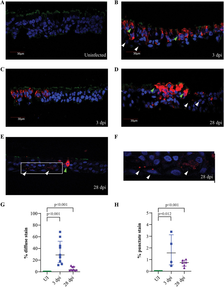
The dynamics of SARS-CoV-2 infection in COVID-19 patients are highly variable, with a subset of patients demonstrating prolonged virus shedding, which poses a significant challenge for disease management and transmission control. In this study, the long-term dynamics of SARS-CoV-2 infection were investigated using a human well-differentiated nasal epithelial cell (NEC) model of infection. NECs were observed to release SARS-CoV-2 virus onto the apical surface for up to 28 days postinfection (dpi), further corroborated by viral antigen staining. Single-cell transcriptome sequencing (sc-seq) was utilized to explore the host response from infected NECs after short-term (3-dpi) and long-term (28-dpi) infection. We identified a unique population of cells harboring high viral loads present at both 3 and 28 dpi, characterized by expression of cell stress-related genes DDIT3 and ATF3 and enriched for genes involved in tumor necrosis factor alpha (TNF-α) signaling and apoptosis. Remarkably, this sc-seq analysis revealed an antiviral gene signature within all NEC cell types even at 28 dpi. We demonstrate increased replication of basal cells, absence of widespread cell death within the epithelial monolayer, and the ability of SARS-CoV-2 to replicate despite a continuous interferon response as factors likely contributing to SARS-CoV-2 persistence. This study provides a model system for development of therapeutics aimed at improving viral clearance in immunocompromised patients and implies a crucial role for immune cells in mediating viral clearance from infected epithelia. IMPORTANCE Increasing medical attention has been drawn to the persistence of symptoms (long-COVID syndrome) or live virus shedding from subsets of COVID-19 patients weeks to months after the initial onset of symptoms. In vitro approaches to model viral or symptom persistence are needed to fully dissect the complex and likely varied mechanisms underlying these clinical observations. We show that in vitro differentiated human NECs are persistently infected with SARS-CoV-2 for up to 28 dpi. This viral replication occurred despite the presence of an antiviral gene signature across all NEC cell types even at 28 dpi. This indicates that epithelial cell intrinsic antiviral responses are insufficient for the clearance of SARS-CoV-2, implying an essential role for tissue-resident and infiltrating immune cells for eventual viral clearance from infected airway tissue in COVID-19 patients.

Keywords: COVID-19; SARS-CoV-2; nasal epithelial cells; single-cell sequencing; viral persistence.


Conflict of interest statement

The authors declare no conflict of interest.

Figures

FIG 1
FIG 1
SARS-CoV-2 replication kinetics in AECs. Apical viral titers from infected NECs (blue) and BECs (orange) at each of the indicated time points (n = 4 to 8), as detected via (A) single-endpoint sampling or (B) repeated sampling of the same apical surface for each time point. Each dot represents a different human-donor-derived AEC. Data are represented as the mean ± standard error of the mean (SEM). P values indicated are derived from Student's t test.
FIG 2
FIG 2
Detection of SARS-CoV-2 viral antigen from infected NECs. (A to F) IF staining for SARS-CoV-2 nucleoprotein (red), DAPI nuclear stain (blue), and βIV-tubulin (green) from (A) uninfected, (B and C) 3-dpi, and (D to F) 28-dpi infected NECs. Green arrows indicate examples of cells with diffuse viral antigen staining, and white arrows indicate cells with punctate staining for viral antigen. Panel F is zoomed in from the indicated image region in panel E to better illustrate punctate focal staining for viral antigen. Representative images obtained from at least 3 different donor-derived NEC inserts processed for IF staining are shown. (G and H) The percentage of cells within NEC structures displaying (G) diffuse viral antigen staining and (H) punctate viral antigen-positive foci at each of the indicated time points. Each dot represents data obtained from a different human donor-derived NEC cross-section. Data are represented as the mean ± SEM. The indicated P values are derived from Student's t test.
FIG 3
FIG 3
Clustering of sc-seq transcriptomes identified 10 airway epithelial cell types within NECs. (A) Pooled sc-seq transcriptomes from uninfected and 3- and 28-dpi NECs clustered and annotated as indicated on the figure. (B) Violin plots illustrating expression of key cell-type markers across the 10 epithelial cell types. (C) Specific expression of cell stress-related genes, DDIT3 and ATF3, from the DDIT3high cell cluster. (D) The percentage change in the proportion of each cell type at 3 and 28 dpi relative to the uninfected NEC samples.
FIG 4
FIG 4
SARS-CoV-2 viral reads detected across multiple epithelial cell types; the DDIT3high cluster consists of heavily infected cells. (A) SARS-CoV-2 viral reads overlaid with clustering of sc-seq transcriptomes derived from uninfected, 3- and 28-dpi NECs. (B) Histograms of viral readsper cell from 3- and 28-dpi NECs. Blue lines indicate the cutoff for demarcating infected versus bystander cells (4 reads per cell), as determined by Otsu’s thresholding. (C) Percentage of cell transcriptomes with >4 and >50 viral reads across each of the epithelial cell types. (D) Sc-seq transcriptomes from the DDIT3high cluster reannotated with transcriptional signatures from the other 9 epithelial cell types in this study and the resulting annotations quantified and displayed as a bar chart on the right panel.
FIG 5
FIG 5
The DDIT3high cluster has reduced IFN response, and higher TNF-σ signaling gene set enrichment. (A) Bubble-plot of the top enriched Hallmark gene sets from differentially expressed genes (DEGs) at 3 dpi compared to uninfected NECs (D3/D0). The color and size of each bubble is proportional to the adjusted P value and the percentage of enriched genes from each gene set, respectively. Only gene-sets with an adjusted P value < 0.05 are shown. (B) Split violin plots illustrating upregulation of key antiviral genes across epithelial cell types at 3 dpi compared to uninfected NECs. The DDIT3high cluster has a relatively lower upregulation of antiviral genes relative to the other cell types. (C) Heat map of the most highly expressed genes in the “TNF-α signaling” (in blue) and “IFN-α response” (in red) Hallmark gene sets across cell transcriptomes derived from 3-dpi NEC samples, arranged according to cell type clusters and sorted for SARS-CoV-2 viral reads (top panel).
FIG 6
FIG 6
A limited number of cells are responsible for producing type I and type III IFN from infected NECs at 3 dpi. (A) Diverse inflammatory mediators produced from various epithelial cell types from 3-dpi infected NECs. (B) Induction of type I and type III IFN genes across epithelial cell types from infected NECs at 3 dpi relative to uninfected NECs (D3/D0). Only IFN genes with detectable expression across the pooled sc-seq data set were included in this analysis. (C) Expression of IFN-β1 and IFN-λ1 across cell transcriptomes from uninfected and 3-dpi infected NEC samples, indicating positive expression of IFN from a very limited subset of cells upon infection.
FIG 7
FIG 7
Broad IFN response observed across cell types from infected NECs at 28 dpi. (A) Bubble plot of IFN-γ and IFN-α response gene set enrichment in DEGs at 28 dpi compared to uninfected NECs (D28/D0). The color and size of each bubble is proportional to the adjusted P value and the percentage of enriched genes from each gene set, respectively. Only gene sets with an adjusted P value of <0.05 are shown. (B) Volcano plot of genes within the goblet 2 cell cluster from infected NECs at 28 dpi compared to uninfected NECs (D28/D0). Significant DEGs with an adjusted P value of <0.05 and log2 fold change (FC) of at least 1.5 are indicated as maroon dots; all other DEGs are indicated as gray dots. The top 15 upregulated genes by log FC are annotated, with genes in the “Hallmark Interferon Alpha response” list annotated in red. (C) Split violin plots illustrating upregulation of key antiviral genes across epithelial cell types at 28 dpi (D3) compared to uninfected NECs (D0), and (D) 3 dpi (D3) compared to 28 dpi (D28) infected NECs.
FIG 8
FIG 8
Elevated CXCL10 levels are observed in basolateral media from infected NECs for up to 28 days postinfection. CXCL10 cytokine concentrations (pg/mL, log10) were assayed from the basolateral media at each of the indicated postinfected time points. Data are shown as the mean ± SEM. Each dot represents a different human-donor-derived NEC. Indicated P values are derived from Student's t test.

References

    1. Qiu X, Nergiz AI, Maraolo AE, Bogoch II, Low N, Cevik M. 2021. The role of asymptomatic and pre-symptomatic infection in SARS-CoV-2 transmission-a living systematic review. Clin Microbiol Infect 27:511–519. doi:10.1016/j.cmi.2021.01.011. - DOI - PMC - PubMed
    1. Meyerowitz EA, Richterman A, Bogoch II, Low N, Cevik M. 2021. Towards an accurate and systematic characterisation of persistently asymptomatic infection with SARS-CoV-2. Lancet Infect Dis 21:e163–e169. doi:10.1016/S1473-3099(20)30837-9. - DOI - PMC - PubMed
    1. Cevik M, Tate M, Lloyd O, Maraolo AE, Schafers J, Ho A. 2021. SARS-CoV-2, SARS-CoV, and MERS-CoV viral load dynamics, duration of viral shedding, and infectiousness: a systematic review and meta-analysis. Lancet Microbe 2:e13–e22. doi:10.1016/S2666-5247(20)30172-5. - DOI - PMC - PubMed
    1. Harvey WT, Carabelli AM, Jackson B, Gupta RK, Thomson EC, Harrison EM, Ludden C, Reeve R, Rambaut A, Consortium C-GU, Peacock SJ, Robertson DL, COVID-19 Genomics UK (COG-UK) Consortium . 2021. SARS-CoV-2 variants, spike mutations and immune escape. Nat Rev Microbiol 19:409–424. doi:10.1038/s41579-021-00573-0. - DOI - PMC - PubMed
    1. Gallo O, Locatello LG, Mazzoni A, Novelli L, Annunziato F. 2021. The central role of the nasal microenvironment in the transmission, modulation, and clinical progression of SARS-CoV-2 infection. Mucosal Immunol 14:305–316. doi:10.1038/s41385-020-00359-2. - DOI - PMC - PubMed

Publication types

Substances