. 2022 Feb 22;13(1):e0368021.
doi: 10.1128/mbio.03680-21. Epub 2022 Jan 18.

Pseudomonas aeruginosa C-Terminal Processing Protease CtpA Assembles into a Hexameric Structure That Requires Activation by a Spiral-Shaped Lipoprotein-Binding Partner

Affiliations

Pseudomonas aeruginosa C-Terminal Processing Protease CtpA Assembles into a Hexameric Structure That Requires Activation by a Spiral-Shaped Lipoprotein-Binding Partner

Hao-Chi Hsu et al. mBio. .

Abstract

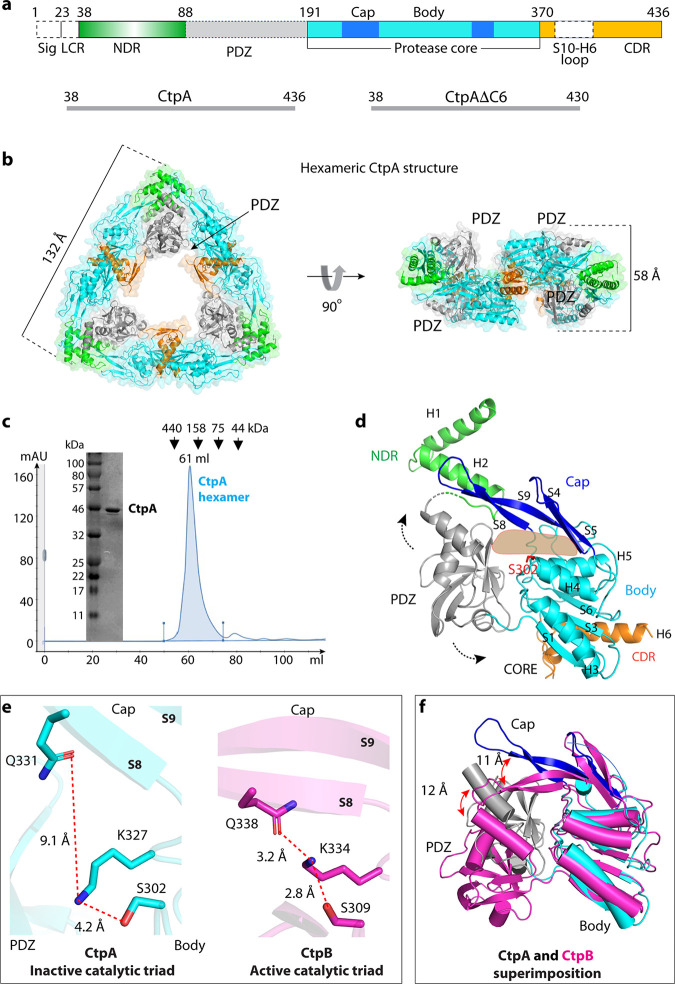
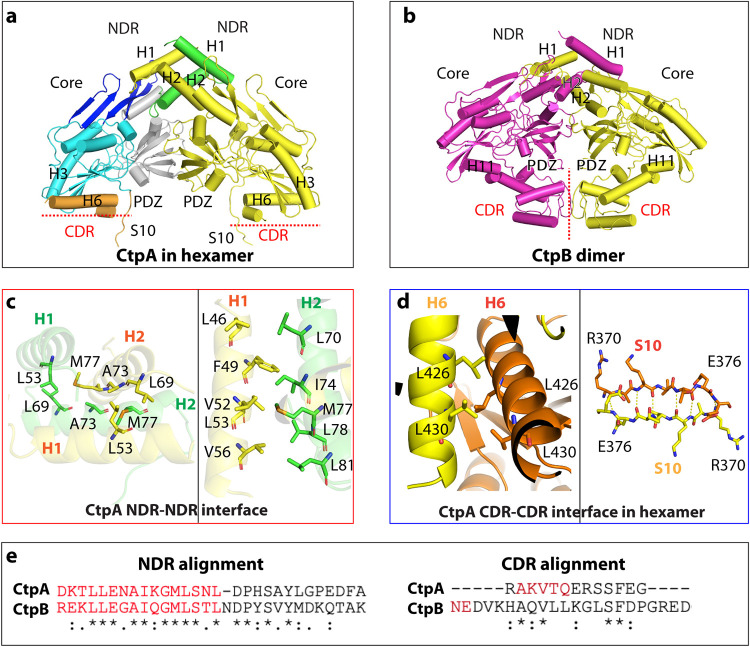
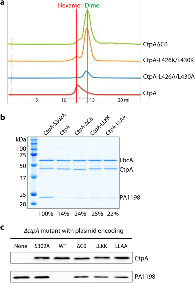
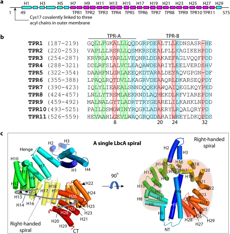
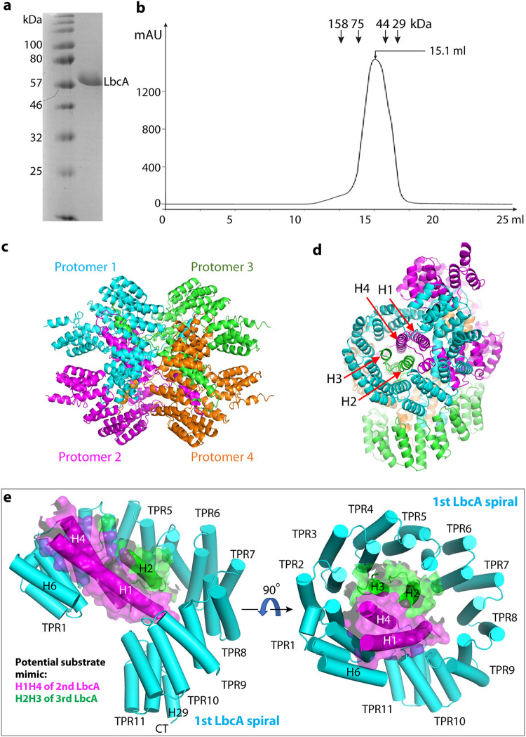
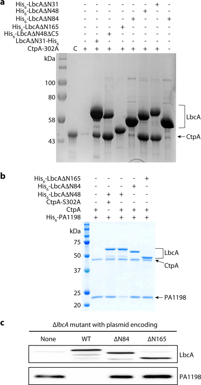
Pseudomonas aeruginosa CtpA is a carboxyl-terminal processing protease that partners with the outer membrane lipoprotein LbcA to degrade at least five cell wall-associated proteins, four of which are cell wall hydrolases. This activity plays an important role in supporting P. aeruginosa virulence in a mouse model of acute pneumonia. However, almost nothing is known about the molecular mechanisms underlying CtpA and LbcA function. Here, we used structural analysis to show that CtpA alone assembles into an inactive hexamer comprising a trimer of dimers, which limits its substrate access and prevents nonspecific degradation. The adaptor protein LbcA is a right-handed open spiral with 11 tetratricopeptide repeats, which might wrap around a substrate to deliver it to CtpA for degradation. By structure-guided mutagenesis and functional assays, we also showed that the interfaces of the CtpA trimer of dimers and an N-terminal helix of LbcA are important for LbcA-mediated substrate degradation by CtpA both in vitro and in vivo. This work improves our understanding of the molecular mechanism of the LbcA-CtpA proteolytic system and reveals some striking differences from the arrangements found in some other bacterial CTPs. IMPORTANCE Carboxyl-terminal processing proteases (CTPs) are found in all three domains of life. In bacteria, some CTPs have been associated with virulence, raising the possibility that they could be therapeutic targets. However, relatively little is known about their molecular mechanisms of action. In Pseudomonas aeruginosa, CtpA supports virulence by working in complex with the outer membrane lipoprotein LbcA to degrade cell wall hydrolases. Here, we report structure-function analyses of CtpA and LbcA, which reveals that CtpA assembles into an inactive hexamer comprising a trimer of dimers. LbcA is monomeric, with the first N-terminal helix important for binding to and activating CtpA, followed by a spiral structure composed of 11 tetratricopeptide repeats, which could wrap around a substrate for delivery to CtpA. This work reveals a unique mutimeric arrangement for a CTP and insight into how the important LbcA-CtpA proteolytic system functions.

Keywords: C-terminal processing protease; Pseudomonas aeruginosa; cell wall; cryo-EM; structural biology.


Conflict of interest statement

The authors declare no conflict of interest.

Figures

FIG 1
FIG 1
Overall structure of CtpA. (a) Top: CtpA domain organization. The dashed lines indicate the disordered regions (PDZ and the S10-H6 connecting loop) that are not resolved in the crystal structure. NDR = N-terminal dimerization region; CDR = C-terminal dimerization region; Sig = signal sequence; LCR = low complexity region. The sequence ranges of the two CtpA constructs used this study are shown in the lower panels. (b) Cartoon and transparent surface views of CtpA hexamer. The domains are colored according to the depiction in panel a. (c) SDS-PAGE analysis and gel filtration profile of CtpA. mAU = milli-absorbance units; kDa = kiloDaltons. (d) A CtpA subunit in cartoon view. Secondary structural elements in CtpA are labeled, except in the PDZ domain. The two dashed arrows indicate the mobile PDZ domain in the CtpA hexamer. The light orange shape indicates the tunnel between the cap and the body region. (e) Comparison of the catalytic triads of P. aeruginosa CtpA and B. subtilis CtpB (PDB ID 4C2E). The S8 and S9 labels refer to β-strands 8 and 9 in the cap region. (f) Superposition of the core domains of inactive P. aeruginosa CtpA (cyan) and active B. subtilis CtpB (magenta). Catalytic Ser-302 in CtpA and Ser-309 in CtpB are in red sticks. Red arrows indicate the lifted-up (CtpA) and clamped-down positions (CtpB) of the PDZ and the cap subdomains.
FIG 2
FIG 2
Different oligomerization modes of P. aeruginosa CtpA and B. subtilis CtpB. (a) CtpA dimer extracted from the CtpA hexamer. (b) B. subtilis CtpB dimer (PDB ID 4C2E). CtpB H11 is the equivalent of CtpA H6. (c) The N-terminal dimerization interface of CtpA involves hydrophobic interactions between two H2s (left) and between H1 and H2 (right). (d) The C-terminal dimerization interface of CtpA involves a short leucine zipper-like interaction between two H6 helices and antiparallel β-sheet formation between two S10s. (e) Alignment of the conserved NDR sequence and divergent CDR between P. aeruginosa CtpA and B. subtilis CtpB.
FIG 3
FIG 3
Full protease activity of CtpA requires C-terminal dimerization region. (a) Elution profiles of wild-type and mutant CtpA proteins. (b) Substrate degradation assay in vitro. His6-PA1198 served as the substrate for the assay. Gels from a single experiment are shown, but the amount of PA1198 degradation is the average from two independent experiments, determined as described in the Materials and Methods section. The number below each lane is the percentage of remaining PA1198 after 3 h, relative to the first lane using the inactive CtpA. (c) Substrate degradation in vivo. Plasmid-encoded protease-dead CtpA(S302A), wild type (WT), ΔC6, L426K/L430K (LLKK), and L426A/L430A (LLAA) were produced in a P. aeruginosa ΔctpA strain. None = empty plasmid vector control. The CtpA proteins and accumulation of the PA1198 substrate were detected by immunoblot analysis with polyclonal antisera.
FIG 4
FIG 4
Overall structure of LbcA. (a) Domain organization of LbcA. The TPR motifs are shown in magenta. TPR = tetratricopeptide repeats. (b) Sequence alignment of the 11 TPRs of LbcA. The signature residues of TPR are marked in the red rectangles. The highly homologous hydrophobic residues are in green boxes, and the highly homologous charged residues are in the blue boxes. (c) The crystal structure of LbcA(ΔN48) contains an N-terminal extension with 4 helices and a C-terminal superhelix composed of 11 TPRs. The thick gray curve in the left panel follows the right-handed spiral feature of the TPRs.
FIG 5
FIG 5
LbcA is a monomer in solution but assembles a tetramer in crystal. (a) Coomassie blue-stained SDS-PAGE gel of the purified of LbcAΔN48. (b) Superdex 200 elution profile of LbcAΔN48. LbcA was eluted from a gel filtration column at a volume corresponding to the monomeric state. (c) LbcA forms an intertwined tetramer in the crystal lattice. The four protomers are individually colored. (d) This LbcA tetramer view shows that the helices H1 and H4 of protomer 2 and helices H2 and H3 of protomer 3 form a 4-helix bundle inside the super helical coil of protomer 1 in the crystal lattice. (e) H1H4 of a second LbcA (magenta cartoon and transparent surface) and H2H3 of a third LbcA (green cartoon and transparent surface) insert into the first LbcA spiral, likely mimicking the substrate binding by the first LbcA spiral.
FIG 6
FIG 6
The LbcA N-terminal extension is essential for the protease activity of CtpA. (a) CtpA pulldown assay using various N-terminal and C-terminal deletion mutants of LbcA. Lane 2 is the CtpA input control (C). Lane 3 is the background binding of CtpA to nickel beads. (b) In vitro substrate (PA1198) degradation assay. (c) Substrate degradation in vivo. Plasmids encoding wild-type LbcA (WT), LbcA(ΔN84), or LbcA(ΔN165) were transformed into a P. aeruginosa ΔlbcA mutant. None = empty plasmid vector control. The LbcA proteins and accumulation of the PA1198 substrate were detected by immunoblot analysis with polyclonal antisera.

References

    1. Vollmer W, Blanot D, de Pedro MA. 2008. Peptidoglycan structure and architecture. FEMS Microbiol Rev 32:149–167. doi:10.1111/j.1574-6976.2007.00094.x. - DOI - PubMed
    1. Pazos M, Peters K, Vollmer W. 2017. Robust peptidoglycan growth by dynamic and variable multi-protein complexes. Curr Opin Microbiol 36:55–61. doi:10.1016/j.mib.2017.01.006. - DOI - PubMed
    1. Vollmer W. 2012. Bacterial growth does require peptidoglycan hydrolases. Mol Microbiol 86:1031–1035. doi:10.1111/mmi.12059. - DOI - PubMed
    1. Park SH, Kim YJ, Lee HB, Seok YJ, Lee CR. 2020. Genetic evidence for distinct functions of peptidoglycan endopeptidases in Escherichia coli. Front Microbiol 11:565767. doi:10.3389/fmicb.2020.565767. - DOI - PMC - PubMed
    1. Singh SK, Parveen S, SaiSree L, Reddy M. 2015. Regulated proteolysis of a cross-link-specific peptidoglycan hydrolase contributes to bacterial morphogenesis. Proc Natl Acad Sci USA 112:10956–10961. doi:10.1073/pnas.1507760112. - DOI - PMC - PubMed

Publication types

LinkOut - more resources