. 2022 Jan 17;24(1):25.
doi: 10.1186/s13075-021-02690-w.

Fibrotic alterations in human annulus fibrosus correlate with progression of intervertebral disc herniation

Affiliations

Fibrotic alterations in human annulus fibrosus correlate with progression of intervertebral disc herniation

A L Castro et al. Arthritis Res Ther. .

Abstract

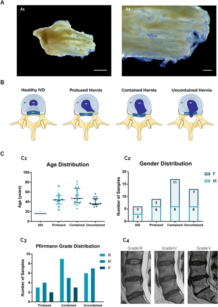
Background: Intervertebral disc (IVD) herniation is characterized by annulus fibrosus failure (AF) in containing the nucleus pulposus (NP). IVD herniation involves cellular and extracellular matrix (ECM) alterations that have been associated with tissue fibrosis, although still poorly investigated.

Methods: Here, fibrotic alterations in human AF were evaluated, by characterizing the herniated ECM. Human AF samples (herniated lumbar IVD (n = 39, age 24-83) and scoliosis controls (n = 6, age 15-21)) were processed for transmission electron microscopy and histological/immunohistochemical analysis of fibrotic markers. Correlations between the fibrotic markers in AF ECM and the degree of NP containment (protused, contained and uncontained) and patients' age were conducted.

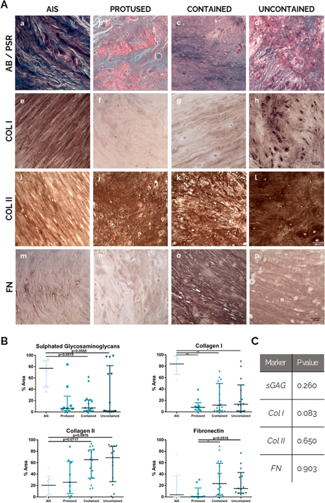
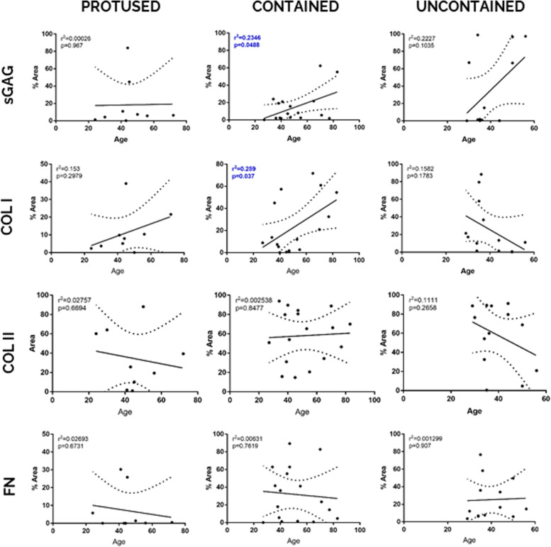
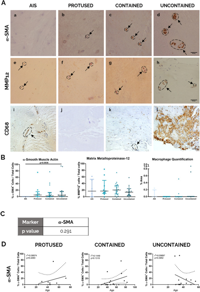
Results: Our results demonstrate that with herniation progression, i.e. loss of NP containment, human AF presents less stained area of sulphated glycosaminoglycans and collagen I, being collagen I fibres thinner and disorganized. On the other hand, fibronectin stained area and percentage of α-smooth muscle actin+ cells increase in human AF, while matrix metalloproteinase-12 (MMP12) production and percentage of macrophages (CD68+ cells) remain constant. These structural and biochemical fibrotic alterations observed in human AF with herniation progression occur independently of the age.

Conclusions: The characterization of human AF here conducted evidence the presence of fibrosis in degenerated IVD, while highlighting the importance of considering the herniation progression stage, despite the patients' age, for a better understanding of the mechanisms behind AF failure and IVD herniation.

Keywords: Annulus fibrosus; Disc herniation; Extracellular matrix; Fibrosis.


Conflict of interest statement

The authors declare that they have no competing interests.

Figures

Fig. 1
Fig. 1
Human AF tissue obtained from LBP patients. A Native hAF tissue after macroscopic separation of IVD samples. Scale bar: A1—5 mm; A2—1 mm. B Schematic representation of human IVD degeneration/herniation. C Clinical data from hAF donors, from scoliosis and LBP patients distributed by hernia containment type (protused, contained, uncontained). C1: age, C2: gender and C3: Pfirrmann grade distribution; C4: representative MRI images of Pfirrmann grade III, IV and V, all in L5-S1 level
Fig. 2
Fig. 2
Human AF matrix ultra-structural characterization with herniation progression (n = 3). A a–d: × 5000 magnification, scale bar: 2 μm; e–l: × 12,000 magnification, scale bar: 1 μm. White arrows point to well organized collagen fibres, while red arrows point to disorganized fibres. B Picro-Sirius Red staining under polarized light, scale bar: 500 μm. C Quantification of collagen fibres per birefringence colour. Kruskal-Wallis test followed by corrected Dunn’s were performed. *p < 0.05; **p < 0.01
Fig. 3
Fig. 3
Human AF matrix biochemical characterization with herniation progression. A Histological/IHC staining for a–d: Alcian Blue/Picro-Sirius Red, scale bar: 100 μm; e–h: Collagen I, scale bar: 100 μm; i–l: Collagen II, scale bar: 100 μm; m–p: Fibronectin, scale bar: 100 μm. B Quantification of each staining per herniation type. Data presented using dot plots, with median and interquartile range. Kruskal-Wallis test followed by corrected Dunn’s were performed. *p < 0.05; **p < 0.01. C Multivariate analysis of interaction of hernia containment level with age for each staining
Fig. 4
Fig. 4
Correlation of expression levels of hAF matrix components with age, within each herniation type. Data presented using dot plots, with 95% confidence intervals and indication of r2 and p value for each linear regression
Fig. 5
Fig. 5
Human AF fibrotic analysis at the cell level with herniation progression. A IHC staining for a–d: α-SMA, scale bar: 50 μm; e–h: MMP12, scale bar: 50 μm; i–l: macrophages (CD68) scale bar: 50 μm. B Quantification of each staining per herniation type. Data presented using dot plots, with median and interquartile range. Kruskal-Wallis test followed by corrected Dunn’s were performed. *p < 0.05; **p < 0.01. C Multivariate analysis of interaction of hernia containment level with age for α-SMA. D Correlation of α-SMA staining quantification with age for each herniation type. Data presented using dot plots with 95% confidence intervals and indication of r2 and p value for each linear regression

References

    1. Chu G, et al. Strategies for annulus fibrosus regeneration: from biological therapies to tissue engineering. Front Bioeng Biotechnol. 2018;6:90. - PMC - PubMed
    1. Inoue N, Espinoza Orías AA. Biomechanics of intervertebral disk degeneration. Orthop Clin North Am. 2011;42(4):487–499. - PMC - PubMed
    1. Cunha C, et al. The inflammatory response in the regression of lumbar disc herniation. Arthritis Res Ther. 2018;20(1):251. - PMC - PubMed
    1. Fardon DF, et al. Lumbar disc nomenclature: version 2.0: recommendations of the combined task forces of the North American Spine Society, the American Society of Spine Radiology and the American Society of Neuroradiology. Spine J. 2014;14(11):2525–2545. - PubMed
    1. Molinos M, et al. Age-correlated phenotypic alterations in cells isolated from human degenerated intervertebral discs with contained hernias. Spine (Phila Pa 1976) 2018;43(5):E274–e284. - PubMed

Publication types

MeSH terms