Peripheral dentinogenic ghost cell tumour
- PMID: 35039348
- PMCID: PMC8768483
- DOI: 10.1136/bcr-2021-245513
Peripheral dentinogenic ghost cell tumour
Abstract
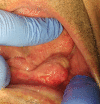
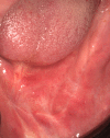
The dentinogenic ghost cell tumour (DGCT) is a rare benign neoplasm, which histologically presents itself as an aberrant keratinisation of the epithelium, ghost cells and dentinoid material. Depending on its location there are two different types of DGCT, central or peripheral, with different clinical characteristics. By 2019, there were only 57 cases of DGCT published: 39 of the central type and 18 of the peripheral type.In this clinical case, the authors describe the case of a 78-year-old man with a painless and slow growing mandibular lump. The diagnosis of peripheral DGCT was made by incisional biopsy and the treatment consisted of radical excision with upper marginal mandibulectomy.The aim of the article is to report a clinical case of a rare pathology and, consequently, to help diagnose and better understand its biological behaviour.
Keywords: dentistry and oral medicine; oral and maxillofacial surgery; pathology.
© BMJ Publishing Group Limited 2022. No commercial re-use. See rights and permissions. Published by BMJ.
Conflict of interest statement
Competing interests: None declared.
Figures


References
-
- El-Naggar AK, Chan JK, Grandis JR. World Health Organization classification of head and neck tumours. 4th ed. Lyon: IARC Press, 2017.
-
- Pinheiro TN, de Souza APF, Bacchi CE, et al. . Dentinogenic ghost cell tumor: a bibliometric review of literature. Jodm 2019;3:9–17. 10.15713/ins.jodm.21 - DOI
-
- Sayd S, Sankar R, Mukund N. Dentinogenic ghost cell Tumour- a review. Acta Scientific Dental Sciences 2019;3.1:130–2.
Publication types
MeSH terms
LinkOut - more resources
Full Text Sources
