. 2022 Jan 17;12(1):828.
doi: 10.1038/s41598-022-04794-y.

Detection of impaired renal allograft function in paediatric and young adult patients using arterial spin labelling MRI (ASL-MRI)

Affiliations

Detection of impaired renal allograft function in paediatric and young adult patients using arterial spin labelling MRI (ASL-MRI)

Tijana Radovic et al. Sci Rep. .

Abstract

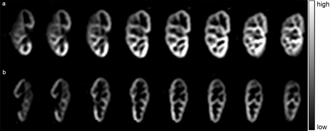
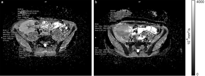
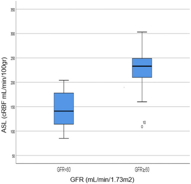
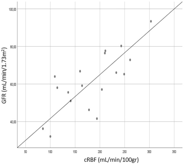
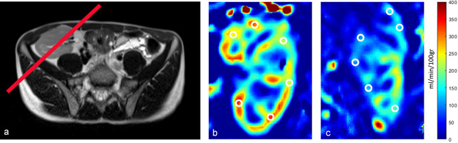
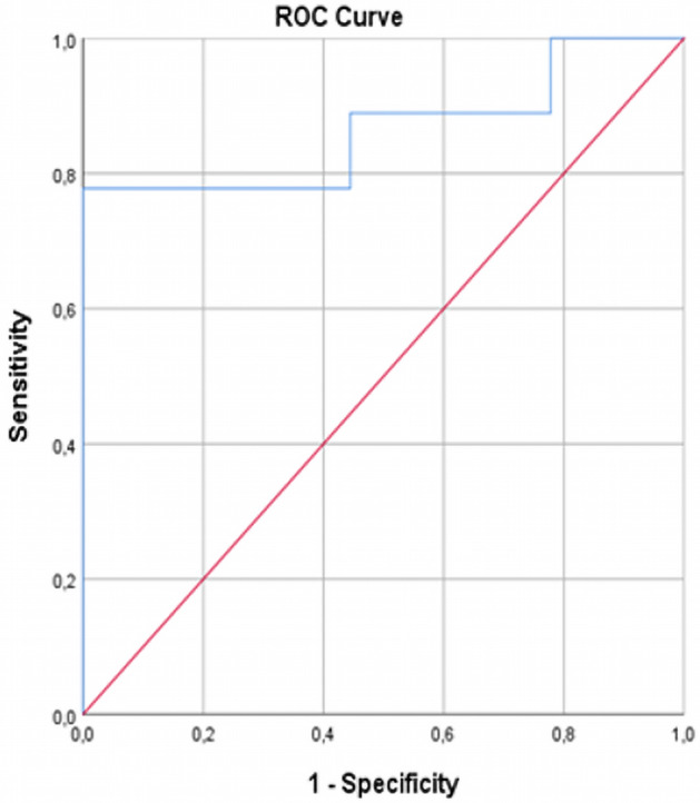
The study aimed to discriminate renal allografts with impaired function by measuring cortical renal blood flow (cRBF) using magnetic resonance imaging arterial spin labelling (ASL-MRI) in paediatric and young adult patients. We included 18 subjects and performed ASL-MRI on 1.5 T MRI to calculate cRBF on parameter maps. cRBF was correlated to calculated glomerular filtration rate (GFR) and compared between patient groups with good (GFR ≥ 60 mL/min/1.73 m2) and impaired allograft function (GFR < 60 mL/min/1.73 m2). Mean cRBF in patients with good allograft function was significantly higher than in patients with impaired allograft function (219.89 ± 57.24 mL/min/100 g vs. 146.22 ± 41.84 mL/min/100 g, p < 0.008), showing a highly significant correlation with GFR in all subjects (r = 0.75, p < 0.0001). Also, the diffusion-weighted imaging (DWI-MRI) apparent diffusion coefficient (ADC) and Doppler measurements of peak-systolic and end-diastolic velocities and the resistive index (PS, ED, RI) were performed and both methods showed no significant difference between groups. ADC implied no correlation with GFR (r = 0.198, p = 0.464), while PS indicated moderate correlation to GFR (r = 0.48, p < 0.05), and PS and ED moderate correlation to cRBF (r = 0.58, p < 0.05, r = 0.56, p < 0.05, respectively). Cortical perfusion as non-invasively measured by ASL-MRI differs between patients with good and impaired allograft function and correlates significantly with its function.


Conflict of interest statement

The authors declare no competing interests.

Figures

Figure 1
Figure 1
Multi-slice perfusion weighted images (PWI) in two representative patients with: (a) good allograft function (GFR ≥ 60 ml/min/100 g) and (b) impaired allograft function (GFR < 60 ml/min/100 g).
Figure 2
Figure 2
Axial ADC maps with representative circular regions of interest (ROI) placed in the renal cortex in: (a) patient with GFR ≥ 60 mL/min/1.73 m2 and (b) patient with GFR ≥ 60 mL/min/1.73 m2 (notice the difference in ADC values of anterior and posterior aspect of the kidney, with moderate diffusion restriction in posterior aspect after intraoperative vascular incident; this DWI appearance resolved after few months from transplantation).
Figure 3
Figure 3
Box plot showing a comparison of perfusion values in patients with GFR < 60 and GFR ≥ 60 mL/min/1.73 m2; an outlier from group 2 (GFR ≥ 60 mL/min/1.73 m2) falling out of the boxplot corresponds to the participant number 10 in Table 1.
Figure 4
Figure 4
Scatter plot showing a positive correlation coefficient between cRBF and GFR; r = 0.75, p < 0.0001.
Figure 5
Figure 5
(a) Positioning of the middle slice; (b) Quantitative Perfusion Map representing cRBF (ml/min/100 g) imaged in a patient with good allograft function (GFR ≥ 60 ml/min/100 g) and (c) Quantitative Perfusion Map representing cRBF (ml/min/100 g) imaged in a patient with impaired allograft function (GFR < 60 ml/min/100 g).
Figure 6
Figure 6
ROC curve of cRBF measured by ASL-MRI for distinguishing allografts with impaired function from allografts with good function.

References

    1. Artz NS, et al. Arterial spin labeling MRI for assessment of perfusion in native and transplanted kidneys. Magn Reson Imaging. 2011;29(1):74–82. doi: 10.1016/j.mri.2010.07.018. - DOI - PMC - PubMed
    1. Lu-Ping L, et al. Evaluation of renal blood flow in chronic kidney disease using arterial spin labeling perfusion magnetic resonance imaging. Kidney Int Rep. 2017;2:36–43. doi: 10.1016/j.ekir.2016.09.003. - DOI - PMC - PubMed
    1. Nery F, et al. Consensus-based technical recommendations for clinical translation of renal ASL MRI. Magn. Reson. Mater. Phys., Biol. Med. 2020;33:141–161. doi: 10.1007/s10334-019-00800-z. - DOI - PMC - PubMed
    1. Nery F, Gordon I, Thomas DL. Non-invasive renal perfusion imaging using arterial spin labeling MRI: Challenges and opportunities. Diagnostics. 2018;8:2. doi: 10.3390/diagnostics8010002. - DOI - PMC - PubMed
    1. Buchanan CE, Cox EF, Francis ST. Evaluation of 2D imaging schemes for pulsed arterial spin labeling of the human kidney cortex. Diagnostics. 2018;8:43. doi: 10.3390/diagnostics8030043. - DOI - PMC - PubMed

MeSH terms