. 2022 Jan 17;12(1):848.
doi: 10.1038/s41598-022-04892-x.

Therapeutic potential of highly functional codon-optimized microutrophin for muscle-specific expression

Affiliations

Therapeutic potential of highly functional codon-optimized microutrophin for muscle-specific expression

Anna V Starikova et al. Sci Rep. .

Abstract

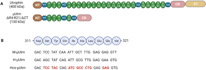
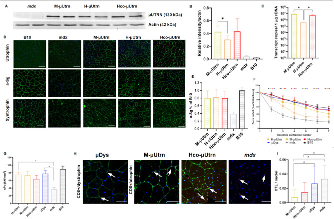
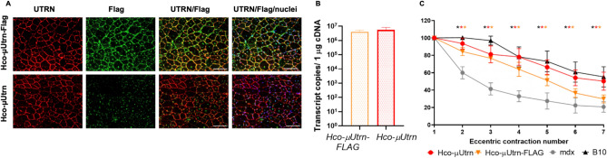
High expectations have been set on gene therapy with an AAV-delivered shortened version of dystrophin (µDys) for Duchenne muscular dystrophy (DMD), with several drug candidates currently undergoing clinical trials. Safety concerns with this therapeutic approach include the immune response to introduced dystrophin antigens observed in some DMD patients. Recent reports highlighted microutrophin (µUtrn) as a less immunogenic functional dystrophin substitute for gene therapy. In the current study, we created a human codon-optimized µUtrn which was subjected to side-by-side characterization with previously reported mouse and human µUtrn sequences after rAAV9 intramuscular injections in mdx mice. Long-term studies with systemic delivery of rAAV9-µUtrn demonstrated robust transgene expression in muscles, with localization to the sarcolemma, functional improvement of muscle performance, decreased creatine kinase levels, and lower immunogenicity as compared to µDys. An extensive toxicity study in wild-type rats did not reveal adverse changes associated with high-dose rAAV9 administration and human codon-optimized µUtrn overexpression. Furthermore, we verified that muscle-specific promoters MHCK7 and SPc5-12 drive a sufficient level of rAAV9-µUtrn expression to ameliorate the dystrophic phenotype in mdx mice. Our results provide ground for taking human codon-optimized µUtrn combined with muscle-specific promoters into clinical development as safe and efficient gene therapy for DMD.


Conflict of interest statement

The authors declare no competing interests.

Figures

Figure 1
Figure 1
Design of µUtrn-coding sequences. (A) Domain structure of full-length utrophin and µUtrn proteins. Domain configuration of µUtrn ∆R4-R21/ΔCT closely resembles microdystrophin R4-R23/ΔCT and consists of an N-terminal actin-binding domain, hinge 1, spectrin-like repeats 1–3, hinge 2, spectrin-like repeat 22, hinge 4, and a cysteine-rich (CR) domain. (B) Alignment of the µUtrn coding sequences used in this study. Fragments of spectrin-like repeat 1 coding sequences are shown for mouse (M-µUtrn), human (H-µUtrn), and codon-optimized human (Hco-µUtrn) µUtrns. Codons optimized for expression in muscle are highlighted in bold red.
Figure 2
Figure 2
Intramuscular administration of rAAV9-µUtrn at dose 2 × 1012 vg/TA muscle leads to robust transgene expression and improves the contractile function of the TA muscle in mdx mice after 2 weeks post injection. (A) Western blot analysis of recombinant μUtrn expression in tibialis anterior (TA) muscles, with α-actin as the loading control. Unprocessed full-length blots are presented in Supplementary Figure S10. (B) Quantitation of µUtrn expression determined via western blot. Data are presented as mean ± SD, * P ≤ 0.05, n = 4/group. (C) Analysis of µUtrn transgene expression via RT-qPCR. (D) Representative images of native and recombinant utrophin as well as α-sarcoglycan (a-Sg) and α1-syntrophin immunofluorescence in TA muscle cross-sections. Nuclei are counterstained with Hoechst 33,342 (blue). Scale bars, 100 µm. (E) Analysis of α-sarcoglycan (a-Sg) expression levels in the sarcolemma of rAAV9-µUtrn-treated muscle in comparison to that in untreated mdx and B10 mice. Data are presented as the mean ± SD, n = 4–10 sections/group. (F) Percentage force drop following 20% eccentric contraction and (G) specific force of TA muscles from mdx mice administered rAAV9-µUtrn as compared to that in vehicle control mice. Values in (C, F), and (G) are presented as the mean ± SEM (N = 8 TA muscles for each group), and statistical significance was set at P ≤ 0.05. (H) Representative images of CD8 + cytotoxic T-lymphocyte immunofluorescence (red dots marked with white arrows) in TA muscle sections. µUtrn and µDys in the sarcolemma were stained in green, while nuclei were stained in blue. Scale bar, 50 μm. (I) Quantitation of CD8 + CTLs in TA muscle sections. CTL number normalized per nuclei, reflecting the cross-sectional area. Values are expressed as the mean ± SD, n = 11–55 sections analyzed per group.
Figure 3
Figure 3
Systemic delivery of rAAV9-Hco-µUtrn at dose 6 × 1014 vg kg−1 for twenty-week studies in mdx mice. (A) Western blot analysis of recombinant µUtrn expression in the heart, tibialis anterior (TA), triceps (Tri), and diaphragm (Dia); loading control, GAPDH. Unprocessed full-length blots are presented in Supplementary Figure S10. (B) RT-qPCR analysis of Hco-µUtrn expression in the heart, gastrocnemius (GAS), triceps, TA, and diaphragm. The dashed line represents the detection levels of negative controls. (C) Representative images of TA muscle cryosection immunofluorescence analysis after rAAV9-Hco-µUtrn administration. Native and recombinant utrophin as well as α-sarcoglycan were colored in green. Nuclei were counterstained with DAPI (blue). Scale bar, 100 µm. (D) Representative images of hematoxylin and eosin (H&E)-stained skeletal muscle, heart, and diaphragm (see Fig. S2 for more organs). Scale bar, 100 µm. (E) Hanging wire test. The maximum hanging time of three trials during a 300-s wire test protocol normalized to mouse mass. (F) Creatine kinase (CK) levels in serum. (G) Percentage force drop following 20% eccentric contraction of TA muscles of mdx mice administered rAAV9-Hco-µUtrn compared to those of vehicle control mice. (H) Western blot analysis of Hco-µUtrn- and µDys-specific antibodies in the sera of treated mice (1:100 dilution). PC—positive control, sample incubated with antibodies against utrophin (Cau22354) and dystrophin (DysB); loading control, GAPDH. Unprocessed full-length blots are presented in Supplementary Figure S10. All values are presented as the mean ± SEM (n = 4 mice), and statistical significance was set at P < 0.05.
Figure 4
Figure 4
Addition of FLAG epitope to Hco-µUtrn does not interfere with the expression, localization, and function of the recombinant protein after intramuscular administration at dose 2 × 1012 vg/TA muscle. (A) Representative images of utrophin (red) and FLAG-epitope (green) immunofluorescence in TA muscles of mdx mice after 2 weeks post rAAV9-Hco-µUtrn and rAAV9-Hco-µUtrn-FLAG administration. Nuclei were counterstained with Hoechst 33,342 (blue). Scale bar, 100 μm. (B) Comparison of µUtrn and µUtrn-FLAG expression level in treated mdx TA muscles, as determined via RT-qPCR. Data are presented in transcript copies per 1 µg cDNA. (C) Percentage force drop following 20% eccentric contractions of rAAV9-Hco-µUtrn and rAAV9-Hco-µUtrn-FLAG-treated mdx TA muscle vs untreated mdx and B10 mouse muscle. All values are presented as the mean ± SEM (n = 8), and statistical significance was set at P ≤ 0.05.
Figure 5
Figure 5
Systemic administration of rAAV9-Hco-µUtrn-FLAG at doses 2 × 1012 and 6 × 1012 vg kg−1 does not cause toxicity in rats after 3 and 14 days post injection. (A) Study design. (B) Transgene expression levels in the diaphragm, heart, tibialis anterior (TA), liver, and gastrocnemius (GAS) of rats, as determined via RT-qPCR. Values are expressed as transcript copies per 1 µg cDNA. The dashed line represents the detection levels of negative controls. (C) Body weight changes in experimental and control animals. (D) Distance traveled in 5 min during the open field test. All values are presented as the mean ± SEM (n = 5/time point). Statistical significance was set at P ≤ 0.05.
Figure 6
Figure 6
Muscle-specific SPc5-12 and MHCK7 promoters drive µUtrn expression and ensure force improvement after intramuscular injection at dose 2 × 1012 vg/TA in mdx mice. (A) Western blot analysis of recombinant µUtrn expression in TA muscles after 2 weeks post injection, with α-actin as the loading control. Unprocessed full-length blots are presented in Supplementary Figure S10. (B) Hco-μUtrn protein levels in rAAV9-Promoter-Hco-µUtrn-treated mdx TA muscles determined via western blot and normalized to α-actin levels. (C) Analysis of µUtrn transgene expression via RT-qPCR. Values are expressed as transcript copies per 1 µg cDNA. The dashed line represents the detection levels of negative controls. (D) Representative images of µUtrn-FLAG (FLAG), α-sarcoglycan (a-Sg), and α1-syntrophin immunofluorescence in TA muscle cross-sections. Nuclei were counterstained with Hoechst 33,342 (blue). Scale bars, 100 μm. (E) Percentage force drop following 20% eccentric contraction and (F) specific force of TA muscles from mdx mice administered rAAV9-µUtrn-FLAG compared to those from vehicle control mice. Maximal isometric force and cross-sectional area of TA muscles are present in Supplementary Figure S11. All values are presented as the mean ± SEM (N = 8 muscles), and statistical significance was set at P ≤ 0.05.

References

    1. Mendell JR, Lloyd-Puryear M. Report of MDA muscle disease symposium on newborn screening for Duchenne muscular dystrophy. Muscle Nerve. 2013;48:21–26. - PubMed
    1. Duan D. Systemic AAV micro-dystrophin gene therapy for duchenne muscular dystrophy. Mol. Ther. 2018;26:2337–2356. - PMC - PubMed
    1. Mendell JR, et al. Dystrophin immunity in Duchenne’s muscular dystrophy. N. Engl. J. Med. 2010;363:1429–1437. - PMC - PubMed
    1. Gramolini AO, Jasmin BJ. Molecular mechanisms and putative signalling events controlling utrophin expression in mammalian skeletal muscle fibres. Neuromuscul. Disord. NMD. 1998;8:351–361. - PubMed
    1. Clerk A, Morris GE, Dubowitz V, Davies KE, Sewry CA. Dystrophin-related protein, utrophin, in normal and dystrophic human fetal skeletal muscle. Histochem. J. 1993;25:554–561. - PubMed

Publication types