Review
. 2022 Jan 18;132(2):e148552.
doi: 10.1172/JCI148552.

Lipid metabolism in autoimmune rheumatic disease: implications for modern and conventional therapies

Affiliations
Review

Lipid metabolism in autoimmune rheumatic disease: implications for modern and conventional therapies

George Robinson et al. J Clin Invest. .

Abstract

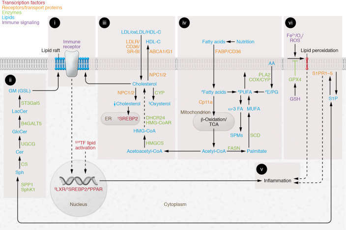
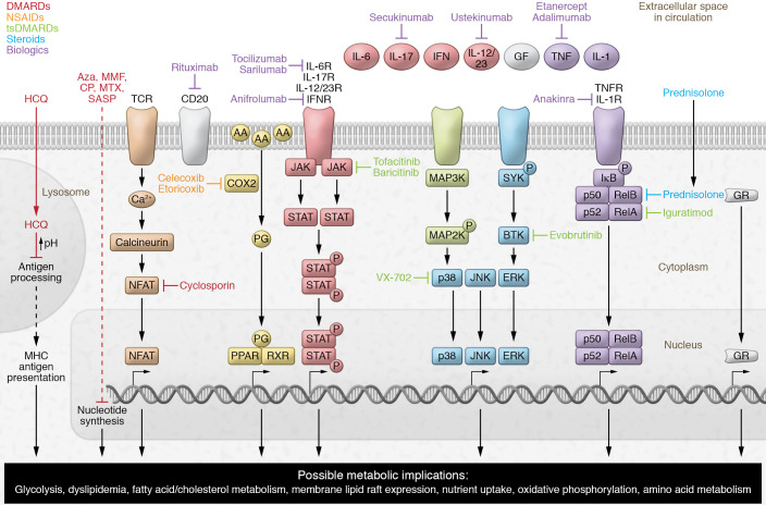
Suppressing inflammation has been the primary focus of therapies in autoimmune rheumatic diseases (AIRDs), including rheumatoid arthritis and systemic lupus erythematosus. However, conventional therapies with low target specificity can have effects on cell metabolism that are less predictable. A key example is lipid metabolism; current therapies can improve or exacerbate dyslipidemia. Many conventional drugs also require in vivo metabolism for their conversion into therapeutically beneficial products; however, drug metabolism often involves the additional formation of toxic by-products, and rates of drug metabolism can be heterogeneous between patients. New therapeutic technologies and research have highlighted alternative metabolic pathways that can be more specifically targeted to reduce inflammation but also to prevent undesirable off-target metabolic consequences of conventional antiinflammatory therapies. This Review highlights the role of lipid metabolism in inflammation and in the mechanisms of action of AIRD therapeutics. Opportunities for cotherapies targeting lipid metabolism that could reduce immunometabolic complications and potential increased cardiovascular disease risk in patients with AIRDs are discussed.


Conflict of interest statement

Conflict of interest: The authors have declared that no conflict of interest exists.

Figures

Figure 1
Figure 1. Summary of lipid metabolism pathways important for inflammation.
(i) Lipid rafts: cholesterol- and glycosphingolipid (GSL)-enriched cell signaling platforms. (ii) De novo GSL biosynthesis: differential expression influences immune receptor–mediated signaling and cell function. (iii) Intracellular cholesterol is regulated by liver X receptor (LXR) and sterol regulatory element–binding protein 2 (SREBP-2) (see v). LXR activation by oxysterols induces cholesterol efflux (ABCA1, ABCG1) and reduces lipid uptake (LDLR, VLDLR, SR-BI, CD36). Niemann-Pick type C1 (NPC1) and NPC2 regulate lysosomal/late endosomal trafficking/recycling of intracellular lipids. SREBP-2 opposes LXR and promotes cholesterol biosynthesis and uptake (HMGCoAR, LDLR). (iv) Nutrition influences fatty acid composition and metabolism. Fatty acids are metabolized to produce energy (ATP) by mitochondrial β-oxidation and TCA. Monounsaturated fatty acids (MUFAs) are synthesized via acetyl-CoA, fatty acid synthase (FASN), and stearoyl-CoA desaturase (SCD). Polyunsaturated fatty acids (PUFAs), diet-derived or biosynthesized in vivo, influence arachidonic acid (AA) metabolism. PUFAs are precursors to triglycerides, phospholipids in plasma membrane, second messengers, hormones, and ketone bodies. Prostaglandins (PGs) are produced following AA release from membrane phospholipids. Downstream PG signaling and eicosanoids have direct metabolic effects on immune cells via PPARs, mediating antiinflammatory effects and modulating LXRs (see v). Omega-3 (ω-3) PUFAs are enzymatically converted to antiinflammatory resolvins (specialized pro-resolving mediators [SPMs]). (v) Lipid metabolism activates transcription factors and influences inflammation via multiple mechanisms (see text). (vi) PUFA phospholipid peroxidation is induced by iron overload and ROS. Products of lipid peroxidation are eliminated via glutathione peroxidase-4 (GPX4); clearance defects induce cell membrane damage and ferroptosis. Sphingosine 1-phosphate (S1P) is derived from membrane phospholipids; activation via binding to S1P receptors (S1PR1–5) initiates immune cell localization to inflammatory sites and T cell differentiation. §, †, and # indicate processes activating LXR, SREBP2, or PPARs, respectively. Other abbreviations: B4GALT5, β-1,4-galactosyltransferase-5; Cer, ceramide; CS, ceramide synthase; DHCR24, 24-dehydrocholesterol reductase; FABPs, fatty acid–binding proteins; GlcCer, glucosylceramide; GM, GM1 ganglioside; GSH, glutathione; HMGCS, 3-hydroxy-3-methylglutaryl–CoA synthase-1; LacCer, lactosylceramide; ox, oxidized; Sph, sphingosine; SphK1, sphingosine kinase-1; SPP1, S1P phosphohydrolase-1; SR-BI, scavenger receptor class B type I; ST3Gal5, ST3 β-galactoside α-2,3-sialyltransferase-5; UGCG, UDP-glucose ceramide glucosyltransferase.
Figure 2
Figure 2. Summary of the mechanisms of action of current therapies used in AIRDs.
Schematic representation summarizing the key mechanistic pathways affected by both traditional and modern therapies used to treat AIRDs, including disease-modifying antirheumatic drugs (DMARDs), target synthetic DMARDs (tsDMARDs), nonsteroidal antiinflammatory drugs (NSAIDs), steroids, and biologics. The majority of these therapeutics result in the modification of immune functions and metabolic pathways through alterations in gene transcription. These pathways provide insight into opportunities for cotherapies to prevent off-target immunometabolic effects. AA, arachidonic acid; Aza, azathioprine; CP, cyclophosphamide; GF, growth factor; GR, glucocorticoid receptor; HCQ, hydroxychloroquine; NF-κB, nuclear factor NF-κB (p50/p52/RelA/RelB/); IκB, inhibitor of κB; MAP2/3K, mitogen-activated protein 2-kinase or 3-kinase; MMF, mycophenolate mofetil; MTX, methotrexate; NFAT, nuclear factor of activated T cells; PG, prostaglandin; R, receptor; RXR, retinoid X receptor; SASP, sulfasalazine; SYK, spleen-associated tyrosine kinase; TCR, T cell receptor.

References

    1. Rhoads JP, et al. Fine tuning of immunometabolism for the treatment of rheumatic diseases. Nat Rev Rheumatol. 2017;13(5):313–320. doi: 10.1038/nrrheum.2017.54. - DOI - PMC - PubMed
    1. Gerriets VA, Rathmell JC. Metabolic pathways in T cell fate and function. Trends Immunol. 2012;33(4):168–173. doi: 10.1016/j.it.2012.01.010. - DOI - PMC - PubMed
    1. Waters LR, et al. Initial B cell activation induces metabolic reprogramming and mitochondrial remodeling. iScience. 2018;5:99–109. doi: 10.1016/j.isci.2018.07.005. - DOI - PMC - PubMed
    1. Galván-Peña S, O’Neill LAJ. Metabolic reprograming in macrophage polarization. Front Immunol. 2014;5:420. - PMC - PubMed
    1. Perl A. Activation of mTOR (mechanistic target of rapamycin) in rheumatic diseases. Nat Rev Rheumatol. 2016;12(3):169–182. doi: 10.1038/nrrheum.2015.172. - DOI - PMC - PubMed

Publication types

MeSH terms