. 2022 Nov 21;145(11):4108-4123.
doi: 10.1093/brain/awac015.

A neuron-to-astrocyte Wnt5a signal governs astrogliosis during HIV-associated pain pathogenesis

Affiliations

A neuron-to-astrocyte Wnt5a signal governs astrogliosis during HIV-associated pain pathogenesis

Xin Liu et al. Brain. .

Abstract

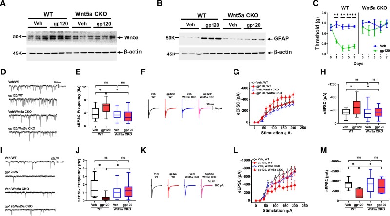
Chronic pain is the most common neurological disorder of HIV patients. Multiple neuropathologies were identified in the pain pathway. Among them is the prominent astrocytic reaction (also know an astrogliosis). However, the pathogenic role and mechanism of the astrogliosis are unclear. Here, we show that the astrogliosis is crucial for the pain development induced by a key neurotoxic HIV protein gp120 and that a neuron-to-astrocyte Wnt5a signal controls the astrogliosis. Ablation of astrogliosis blocked the development of gp120-induced mechanical hyperalgesia, and concomitantly the expression of neural circuit polarization in the spinal dorsal horn. We demonstrated that conditional knockout of either Wnt5a in neurons or its receptor ROR2 in astrocytes abolished not only gp120-induced astrogliosis but also hyperalgesia and neural circuit polarization. Furthermore, we found that the astrogliosis promoted expression of hyperalgesia and NCP via IL-1β regulated by a Wnt5a-ROR2-MMP2 axis. Our results shed light on the role and mechanism of astrogliosis in the pathogenesis of HIV-associated pain.

Keywords: HIV-1 gp120; Wnt; astrocyte; neural circuit; pain.


Conflict of interest statement

The authors report no competing interests.

Figures

Figure 1
Figure 1
Ablation of astrogliosis blocks gp120-induced hyperalgesia and NCP in the SDH. (A) Immunoblotting analysis of spinal GFAP proteins. GCV administration (5 mg/kg at Days 0 and 1, i.t.) ablated astrogliosis inducedby gp120 (4 μg/kg at Days 0, 1, 3 and 5, i.t.) in GFAP-TK transgenic but not in wild-type (WT) mice. Tissues were collected at Day 7 and GFAP protein levels were determined by immunoblotting analysis. (B) Von Frey tests. GCV administration blocked the expression of gp120-induced mechanical hyperalgesia in the GFAP-TK transgenic but not wild-type mice (five mice/group; **P < 0.01). (CH) Effect of ablation of astrogliosis on sEPSCs and eEPSCs of non-tonic firing neurons in the SDH of GFAP-TK transgenic mice. Whole cell patch recording was performed on spinal slices prepared from GFAP-TK transgenic mice treated with gp120 and/or GCV (at Day 7 in B). (C) Non-tonic firing pattern. (D) Representative traces of sEPSCs. (E) Statistical analysis of sEPSC frequency shown in D. gp120 significantly increased sEPSC frequency, and GCV abolished the increase. GCV did not affect the basal level of sEPSC frequency. Veh/Veh: 21/3 (cells/animals); veh/gp120: 20/3; GCV/Veh: 29/4; GCV/gp120: 47/4. (F) Representative traces of eEPSCs. (G). Stimulus-response curve of eEPSC. gp120 increased eEPSC amplitudes, and GCV abolished the increase. GCV did not affect the basal level of eEPSC amplitudes. Veh/Veh: 24/3 (cells/animals); veh/gp120: 24/4; GCV/Veh: 35/3; GCV/gp120: 24/4. (H) Statistical analysis of eEPSC amplitudes evoked by 200 µA stimulation shown in G. (IN) Effect of ablation of astrogliosis on sEPSCs and eEPSCs of tonic firing neurons in the SDH of GFAP-TK transgenic mice. (I) Tonic firing pattern. (J) Representative traces of sEPSCs. (K) Statistical analysis of sEPSC frequency shown in J. gp120 significantly decreased sEPSC frequency. Although GCV treatment by itself also decreased sEPSC frequency, gp120 did not cause further decrease after GCV treatment. Veh/Veh: 13/3 (cells/animals); veh/gp120: 10/3; GCV/Veh: 20/3; GCV/gp120: 26/4. (L) Representative traces of eEPSCs. (M). Stimulus-response curve of eEPSC. gp120 decreased eEPSC amplitudes. GCV also decreased eEPSC amplitudes by itself, but gp120 did not further decrease the amplitudes after GCV administration. Veh/Veh: 14/3 (cells/animals); veh/gp120: 12/4; GCV/Veh: 20/3; GCV/gp120: 16/4. (N) Statistical analysis of eEPSC amplitudes evoked by 200 µA stimulation shown in M. *P < 0.05; not significant (ns), P > 0.05.
Figure 2
Figure 2
Wnt5a CKO in neurons abolishes gp120-induced astrogliosis, hyperalgesia and NCP. (A) Immunoblotting analysis showed that neuronal Wnt5a CKO abolished Wnt5a upregulation induced by gp120. Tissues were collected at Day 7. (B) Immunoblotting analysis of spinal GFAP proteins indicated that neuronal Wnt5a CKO blocked astrogliosis induced by gp120 (described in Fig. 1A) Tissues were collected at Day 7. (C) Von Frey tests showed that neuronal Wnt5a CKO blocked the expression of gp120-induced mechanical hyperalgesia (five mice/group; **P < 0.01). (DH) Effect of neuronal Wnt5a CKO on the sEPSC frequency and eEPSC amplitude of non-tonic firing neurons in the SDH. (D) Representative traces of sEPSCs. (E) Statistical analysis of sEPSC frequency shown in D. Neuronal Wnt5a CKO blocked gp120-induced increase of sEPSC frequency of non-tonic firing neurons in the SDH. Whole cell patch recording was performed on spinal slices as in Fig. 1. gp120 significantly increased sEPSC frequency in wild-type (WT) but not in the Wnt5a CKO mutant mice. Veh/WT: 19/3 (cells/animals); WT/gp120: 21/4; Wnt5a CKO/Veh: 57/5; Wnt5a CKO/gp120: 40/4. (F) Representative traces of eEPSCs. eEPSC amplitudes were recorded on spinal slices prepared as in D. (G) Stimulus-response curve of eEPSC. gp120 increased eEPSC amplitudes in wild-type but not in the Wnt5a CKO mice. Veh/WT: 24/3 (cells/animals); WT/gp120: 24/4; Wnt5a CKO/Veh: 55/5; Wnt5a CKO/gp120: 53/4. (H) Statistical analysis of eEPSC amplitudes evoked by 200 µA stimulation shown in G. (IM) Effect of the neuronal Wnt5a CKO on sEPSCs and eEPSCs of tonic firing neurons in the SDH. (I) Representative traces of eEPSCs. (J). Statistical analysis of sEPSC frequency shown in I. gp120 significantly decreased sEPSC frequency in wild-type but not in the Wnt5a CKO mutant mice. Veh/WT: 12/3 (cells/animals); WT/gp120: 15/4; Wnt5a CKO/Veh: 28/5; Wnt5a CKO/gp120: 9/4. (K) Representative traces of eEPSCs. (L) Stimulus-response curve of eEPSC. gp120 decreased eEPSC amplitudes in the wild-type but not in the Wnt5a CKO mutant mice. Veh/WT: 8/3 (cells/animals); WT/gp120: 10/3; Wnt5a CKO/Veh: 24/3; Wnt5a CKO/gp120: 10/3. (M) Statistical analysis of eEPSC amplitudes evoked by 200 µA stimulation shown in L. *P < 0.05; ns, P > 0.05.
Figure 3
Figure 3
Astrocytic ROR2 CKO diminishes gp120-induced astrogliosis, hyperalgesia and NCP. (A) Immunoblotting analysis of spinal GFAP protein. (B) Statistical analysis of immunoblotting shown in A. Astrocytic ROR2 CKO significantly impaired astrogliosis induced by gp120 (described in Fig. 1A). Tissues were collected at Day 7. (C) Von Frey tests showed that astrocytic ROR2 CKO inhibited the expression of gp120-induced mechanical hyperalgesia. five mice/group; *P < 0.05; **P < 0.01. (DH) Effect of astrocytic ROR2 CKO on the sEPSC frequency and eEPSC amplitude of non-tonic firing neurons in the SDH. (D) Representative traces of sEPSCs. (E) Statistical analysis of sEPSC frequency shown in C. Astrocytic ROR2 CKO inhibited gp120-induced increase of sEPSC frequency of non-tonic firing neurons in the SDH. gp120 significantly increased sEPSC frequency in wild-type but not in the ROR2 CKO mutant mice. Veh/WT: 32/3 (cells/animals); WT/gp120: 18/5; ROR2 CKO/Veh: 45/3; ROR2 CKO/gp120: 32/4. (F) Representative traces of eEPSCs. eEPSC amplitudes were recorded on spinal slices prepared as in C. (G) Stimulus-response curve of eEPSC. gp120 increased eEPSC amplitudes in wild-type but not in the ROR2 CKO mice. Veh/WT: 24/3 (cells/animals); WT/gp120: 36/4; ROR2 CKO/Veh: 34/3; ROR2 CKO/gp120: 38/3. (H) Statistical analysis of eEPSC amplitudes evoked by 200 µA stimulation shown in F. (IM) Effect of astrocytic ROR2 CKO on the sEPSC frequency and eEPSC amplitude of tonic firing neurons in the SDH. (I) Representative traces of sEPSCs. (J) Statistical analysis of sEPSC frequency shown in H. gp120 significantly decreased sEPSC frequency in wild-type but not in the ROR2 CKO mutant mice. Veh/WT: 19/3 (cells/animals); WT/gp120: 24/4; ROR2 CKO/Veh: 22/3; ROR2 CKO/gp120: 31/3. (K) Representative traces of eEPSCs. (L) Stimulus-response curve of eEPSC. gp120 decreased eEPSC amplitudes in the wild-type but not in the ROR2 CKO mutant mice. Veh/WT: 8/3 (cells/animals); WT/gp120: 11/3; ROR2 CKO/Veh: 16/3; ROR2 CKO/gp120: 18/3. (M) Statistical analysis of eEPSC amplitudes evoked by 200 µA stimulation shown in K. *P < 0.05; ns, P > 0.05.
Figure 4
Figure 4
Wnt5a-ROR2 signalling facilitates gp120-induced hyperalgesia and NCP by upregulating IL-1b in reactive astrocytes. (A) Immunostaining of GFAP and IL-1β in the SDH from mice treated with gp120 as described in Fig. 1A. The grey matter is outlined with a white dotted line. Blue and red boxes indicate the regions where white matter astrocytes and grey matter astrocytes are exemplified with higher-power images (in lower panels), respectively. IL-1β signals were mainly observed in both white-matter and grey-matter astrocytes (arrows). (B) Immunoblotting analysis showed that ablation of reactive astrocytes in the GFAP-TK mice diminished gp120-induced spinal IL-1β up-regulation. (C) Neuronal Wnt5a CKO blocked gp120-induced spinal IL-1β upregulation. Spinal cords were collected for immunoblotting from mice at Day 7 after gp120 administration. (D) Astrocytic ROR2 CKO partially but significantly inhibited gp120-induced spinal IL-1β upregulation.
Figure 5
Figure 5
IL-1Ra blocks gp120-induced pain and NCP. (A) IL-1β receptor antagonist IL-1Ra (4 μg/kg/day, three times, intrathecal) blocked the expression of mechanical hyperalgesia induced by gp120 (described in Fig. 1A), measured by von Frey tests (five mice/group; ns, P > 0,05; **P < 0.01). (BF) Effects of IL-1Ra on sEPSC frequency and eEPSC amplitude in non-tonic firing SDH neurons. (B) Representative traces of sEPSCs. (C) Statistical analysis of sEPSC frequency shown in B. IL-1Ra impaired gp120-induced increase of sEPSC frequency in non-tonic firing SDH neurons on spinal slices from mice at Day 7 as shown in E. [Veh/Veh: 24/3 (cells/animals); veh/gp120: 50/5; IL-1Ra/Veh: 24/5; IL-1Ra/gp120: 38/3]. (D) Representative traces of eEPSCs. eEPSC amplitudes were recorded on spinal slices prepared as in B. (E) Stimulus-response curve of eEPSC. IL-1Ra diminished gp120-induced increase of eEPSC amplitudes of non-tonic firing neurons in the SDH. (F) Statistical analysis of eEPSC amplitudes evoked by 200 µA stimulation shown in E [Veh/Veh: 24/3 (cells/animals); veh/gp120: 11/3; IL-1Ra/Veh: 19/3; IL-1Ra/gp120: 38/3]. (GK) Effects of IL-1Ra on sEPSC frequency and eEPSC amplitude in tonic firing SDH neurons. (G) Representative traces of sEPSCs. (H) Statistical analysis of sEPSC frequency shown in G. IL-1Ra blocked gp120-induced decrease of sEPSC frequency in SDH inhibitory neurons of GAD67-GFP transgenic mice [Veh/Veh: 30/3 (cells/animals); veh/gp120: 16/3; IL-1Ra/Veh: 35/3; IL-1Ra/gp120: 16/3]. (I) Representative traces of eEPSCs. eEPSC amplitudes were recorded on spinal slices prepared as in G. (J) Stimulus-response curve of eEPSC. IL-1Ra abolished gp120-induced decrease of eEPSC amplitudes of SDH inhibitory neurons. (K) Statistical analysis of eEPSC amplitudes evoked by 200 µA stimulation shown in J [Veh/Veh: 42/4 (cells/animals); veh/gp120: 16/3; IL-1Ra/Veh: 31/3; IL-1Ra/gp120: 16/3]. *P < 0.05; ns, P > 0.05.
Figure 6
Figure 6
MMP2 is critical for gp120 to induce IL-1β and hyperalgesia. (A) MMP inhibitor GM6001 (4 μg/kg, daily, first 4 days, intrathecal) blocked the expression of mechanical hyperalgesia induced by gp120. n = 5 mice/group. **P < 0.01: ns = not significant. (B) Immunoblotting analysis showed that GM6001, but not YVAD, blocked spinal IL-1β increase induced by gp120. Tissues were collected at Day 7. (C) gp120 induced MMP2 but not MMP9 in the mouse spinal cord (Veh represent control group. n = 5 mice/group). (D) gp120-induced MMP2 protein increase was blocked by MMP2-specific siRNA. Shown are summary data of immunoblotting analysis. n = 5 mice/group. (E) gp120-induced IL-1β was blocked by MMP2-specific siRNA. Shown are summary data of immunoblotting analysis. n = 5 mice/group. (F) MMP2 siRNA, but not MMP9 siRNA, impaired the expression of gp120-induced mechanical hyperalgesia (n = 5 mice/group). *P < 0.05; **P < 0.01: ns = not significant.
Figure 7
Figure 7
Wnt5a CKO in neurons or ROR2 CKO in astrocytes block gp120-induced MMP2 up-regulation in the SDH. (A) Neuronal Wnt5a CKO abolished gp120-induced spinal MMP2 upregulation. n = 5 mice/group. (B) Astrocytic ROR2 CKO significantly impaired gp120-induced spinal MMP2 protein increase. Tissues were collected at Day 7. n = 5 mice/group; *P < 0.05; **P < 0.01. (C) A model. The neuron-to-astrocyte Wnt5a-ROR2 signalling pathway regulates the development of gp120-induced pain via controlling astrogliosis and astrocytic IL-1β activation. IL-1β then induces NCP and pain.

Similar articles

Cited by

References

    1. Parker R, Stein DJ, Jelsma J. Pain in people living with HIV/AIDS: a systematic review. J Int AIDS Soc. 2014;17(1):18719–18719. - PMC - PubMed
    1. Madden VJ, Parker R, Goodin BR. Chronic pain in people with HIV: a common comorbidity and threat to quality of life. Pain Manag. 2020;10(4):253–260. - PMC - PubMed
    1. Lawson E, Sabin C, Perry N, et al. . Is HIV painful? An epidemiologic study of the prevalence and risk factors for pain in HIV-infected patients. Clin J Pain. 2015;31(9):813–819. - PMC - PubMed
    1. Sabin CA, Harding R, Bagkeris E, et al. . Pain in people living with HIV and its association with healthcare resource use, well being and functional status. AIDS. 2018;32(18):2697–2706. - PubMed
    1. Harding R, Sherr L. Comment on ‘Pain in people living with HIV/AIDS: a systematic review (Parker et al. 2014)’. J Int AIDS Soc. 2014;17(1):19096–19096. - PMC - PubMed

Publication types