

Variability of EEG electrode positions and their underlying brain regions: visualizing gel artifacts from a simultaneous EEG-fMRI dataset
- PMID: 35040596
- PMCID: PMC8865144
- DOI: 10.1002/brb3.2476
Variability of EEG electrode positions and their underlying brain regions: visualizing gel artifacts from a simultaneous EEG-fMRI dataset
Abstract
Introduction: We investigated the between-subject variability of EEG (electroencephalography) electrode placement from a simultaneously recorded EEG-fMRI (functional magnetic resonance imaging) dataset.
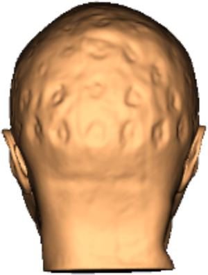
Methods: Neuro-navigation software was used to localize electrode positions, made possible by the gel artifacts present in the structural magnetic resonance images. To assess variation in the brain regions directly underneath electrodes we used MNI coordinates, their associated Brodmann areas, and labels from the Harvard-Oxford Cortical Atlas. We outline this relatively simple pipeline with accompanying analysis code.
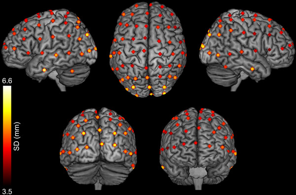
Results: In a sample of 20 participants, the mean standard deviation of electrode placement was 3.94 mm in x, 5.55 mm in y, and 7.17 mm in z, with the largest variation in parietal and occipital electrodes. In addition, the brain regions covered by electrode pairs were not always consistent; for example, the mean location of electrode PO7 was mapped to BA18 (secondary visual cortex), whereas PO8 was closer to BA19 (visual association cortex). Further, electrode C1 was mapped to BA4 (primary motor cortex), whereas C2 was closer to BA6 (premotor cortex).
Conclusions: Overall, the results emphasize the variation in electrode positioning that can be found even in a fixed cap. This may be particularly important to consider when using EEG positioning systems to inform non-invasive neurostimulation.
Keywords: EEG cap | gel artifact; EEG-fMRI; TMS neuro-navigation; electrode positions.
© 2022 The Authors. Brain and Behavior published by Wiley Periodicals LLC.
Figures
References
-
- Beynel, L. , Appelbaum, L. G. , Luber, B. , Crowell, C. A. , Hilbig, S. A. , Lim, W. , Nguyen, D. , Chrapliwy, N. A. , Davis, S. W. , Cabeza, R. , Lisanby, S. H. , & Deng, Z.‐D. (2019). Effects of online repetitive transcranial magnetic stimulation (rTMS) on cognitive processing: A meta‐analysis and recommendations for future studies. Neuroscience & Biobehavioral Reviews, 107, 47–58. 10.1016/j.neubiorev.2019.08.018 - DOI - PMC - PubMed
-
- Bhutada, A. S. , Sepúlveda, P. , Torres, R. , Ossandón, T. , Ruiz, S. , & Sitaram, R. (2020). Semi‐automated and direct localization and labeling of EEG electrodes using MR structural images for simultaneous fMRI‐EEG. Frontiers in Neuroscience, 14, 1134. 10.3389/fnins.2020.558981 - DOI - PMC - PubMed
Publication types
MeSH terms
Grants and funding
LinkOut - more resources
Full Text Sources
Other Literature Sources
Medical
Miscellaneous
