Review
. 2022 May;407(3):957-964.
doi: 10.1007/s00423-022-02436-5. Epub 2022 Jan 18.

Endoscopic vacuum therapy in the upper gastrointestinal tract: when and how to use it

Affiliations
Review

Endoscopic vacuum therapy in the upper gastrointestinal tract: when and how to use it

Christian A Gutschow et al. Langenbecks Arch Surg. 2022 May.

Abstract

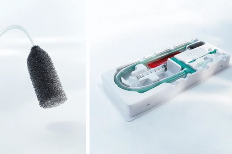
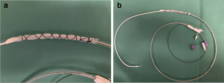
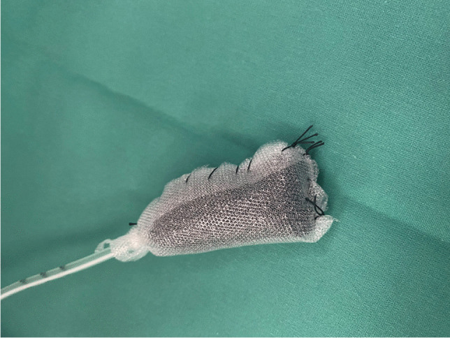
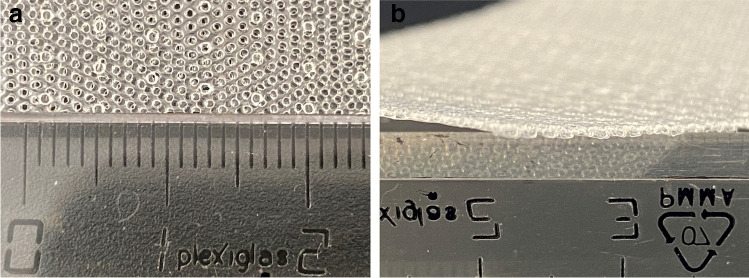
Background: Endoscopic vacuum therapy (EVT) has emerged as a novel treatment option for upper gastrointestinal wall defects. The basic principle of action of EVT entails evacuation of secretions, removal of wound debris, and containment of the defect. Furthermore, there is increasing evidence that EVT reduces interstitial edema, increases oxygen saturation, and promotes tissue granulation and microcirculation. Various devices, such as macroporous polyurethane sponge systems or open-pore film drains, have been developed for specific indications. Depending on the individual situation, EVT devices can be placed in- or outside the intestinal lumen, as a stand-alone procedure, or in combination with surgical, radiological, and other endoscopic interventions.

Purpose: The aim of this narrative review is to describe the current spectrum of EVT in the upper gastrointestinal tract and to assess and summarize the related scientific literature.

Conclusions: There is growing evidence that the efficacy of EVT for upper GI leakages exceeds that of other interventional treatment modalities such as self-expanding metal stents, clips, or simple drainages. Owing to the promising results and the excellent risk profile, EVT has become the therapy of choice for perforations and anastomotic leakages of the upper gastrointestinal tract in many centers of expertise. In addition, recent clinical research suggests that preemptive use of EVT after high-risk upper gastrointestinal resections may play an important role in reducing postoperative morbidity.

Keywords: Anastomotic leakage; Endoscopic vacuum therapy; Esophageal perforation; Esophagectomy; Gastrectomy; Negative pressure therapy.


Conflict of interest statement

CAG received a research grant from B. Braun Melsungen AG, Melsungen, Germany. The other authors declare that they have no conflict of interest.

Figures

Fig. 1
Fig. 1
Small nodules of granulation tissue (micro-deformation) are visible after removal of an EVT device. The extent of micro-deformation depends on various factors such as the porosity of the connecting material, the intensity of the negative pressure, and the properties of the wound surface
Fig. 2
Fig. 2
The EsoSPONGE® device (B.Braun) consists of a low-density macroporous PU foam fitted to a pressure-resistant plastic tube. It is supplied with a specifically designed insertion set using an overtube
Fig. 3
Fig. 3
a and b A self-manufactured open-pore foil drainage for use in the upper GI tract. The perforated part of a three-lumen jejunal feeding and gastric decompression tube (Freka® Trelumina, Fresenius Kabi Deutschland GmbH, Bad Homburg, Germany) is wrapped with a double-layer open-pore film (Suprasorb® CNP Drainage Film, Lohmann & Rauscher International GmbH & Co, Rengsdorf, Germany).
Fig. 4
Fig. 4
A PU foam drain coated with an open-pored film (Suprasorb® CNP Drainage Film, Lohmann & Rauscher International GmbH & Co, Rengsdorf, Germany) to reduce device ingrowth into vulnerable tissues
Fig. 5
Fig. 5
a and b Double-layered open-pored film (Suprasorb® CNP Drainage Film, Lohmann & Rauscher International GmbH & Co, Rengsdorf, Germany) as used by our team for open-pore foil drains.
Fig. 6
Fig. 6
a and b The VacStent® (VAC Stent Medtec AG, Steinhausen, Switzerland) is specifically designed for EVT and combines a cylindrical PU foam with a covered SEMS. b shows a contrast radiography of patient with perforation of the esophago-gastric junction treated with a VacStent®
Fig. 7
Fig. 7
A PU sponge is cut to fit and placed extraluminally/intracavitary with a small extension into the esophageal lumen to drain the abscess cavity and seal the dehiscence, but also to allow free flow of saliva

References

    1. Morykwas MJ, et al. Vacuum-assisted closure: state of basic research and physiologic foundation. Plast Reconstr Surg. 2006;117(7 Suppl):121S–126S. doi: 10.1097/01.prs.0000225450.12593.12. - DOI - PubMed
    1. Weidenhagen R, et al. Endoscopic vacuum-assisted closure of anastomotic leakage following anterior resection of the rectum: a new method. Surg Endosc. 2008;22(8):1818–1825. doi: 10.1007/s00464-007-9706-x. - DOI - PubMed
    1. Loske G, Muller C. Vacuum therapy of an esophageal anastomotic leakage—a case report. Zentralbl Chir. 2009;134(3):267–270. doi: 10.1055/s-0028-1098764. - DOI - PubMed
    1. Loske G. Endoscopic negative pressure therapy of the upper gastrointestinal tract. Chirurg. 2019;90(Suppl 1):1–6. doi: 10.1007/s00104-018-0727-x. - DOI - PMC - PubMed
    1. Sartre J-P (1943) L’Etreet le Néant; Essai D’Ontologie Phenomenologique. Gallimard Paris

LinkOut - more resources