. 2022 Mar 15;66(3):e0194321.
doi: 10.1128/AAC.01943-21. Epub 2022 Jan 18.

Selective Estrogen Receptor Modulators Limit Alphavirus Infection by Targeting the Viral Capping Enzyme nsP1

Affiliations

Selective Estrogen Receptor Modulators Limit Alphavirus Infection by Targeting the Viral Capping Enzyme nsP1

Rajat Mudgal et al. Antimicrob Agents Chemother. .

Abstract

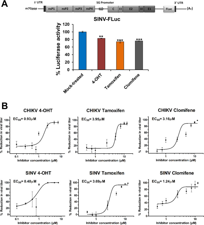
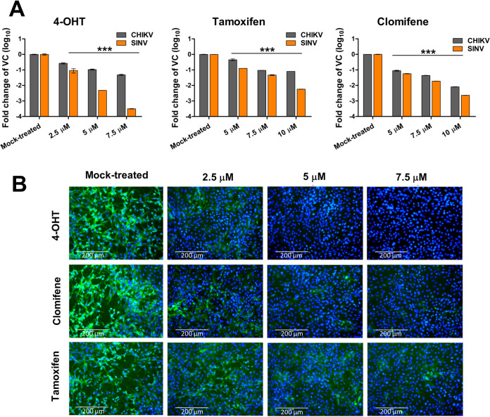
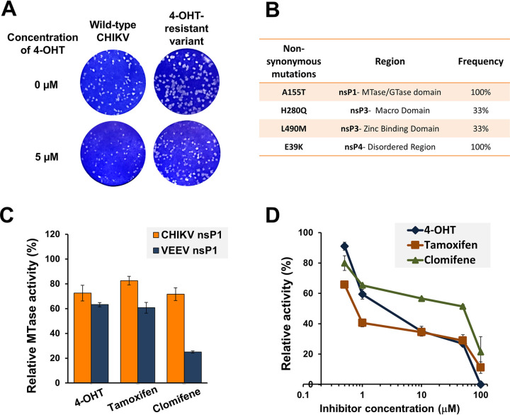
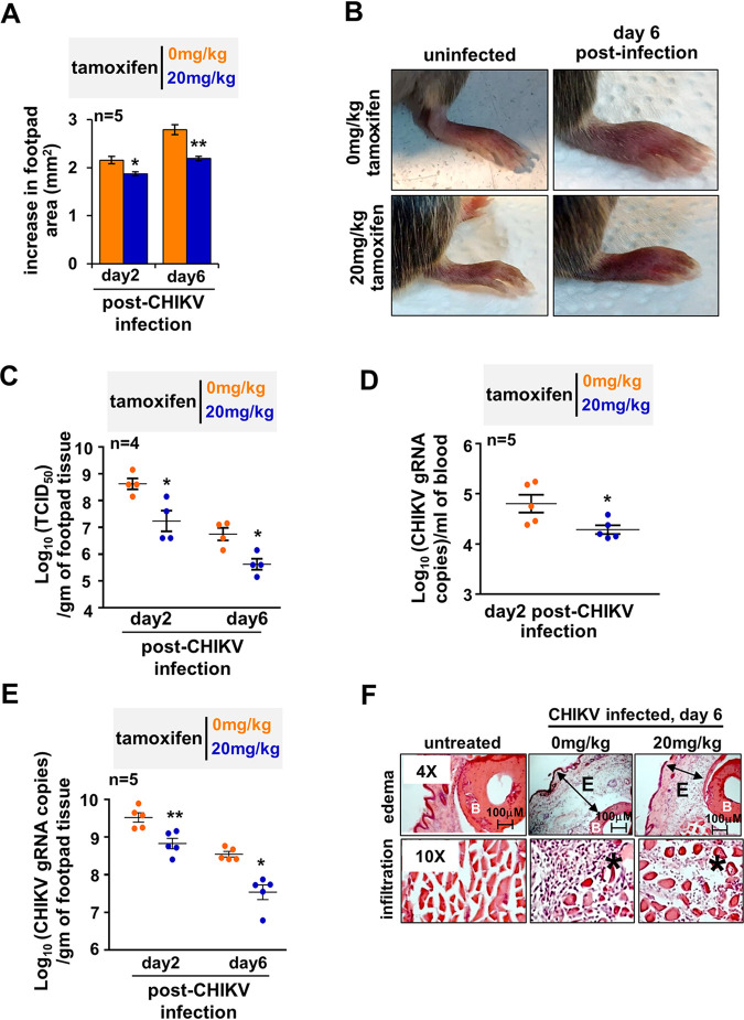
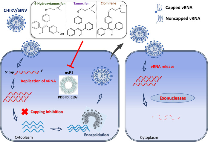
Alphaviruses cause animal or human diseases that are characterized by febrile illness, debilitating arthralgia, or encephalitis. Selective estrogen receptor modulators (SERMs), a class of FDA-approved drugs, have been shown to possess antiviral activities against multiple viruses, including hepatitis C virus, Ebola virus, dengue virus, and vesicular stomatitis virus. Here, we evaluated three SERM compounds, namely, 4-hydroxytamoxifen, tamoxifen, and clomifene, for plausible antiviral properties against two medically important alphaviruses, chikungunya virus (CHIKV) and Sindbis virus (SINV). In cell culture settings, these SERMs displayed potent activity against CHIKV and SINV at nontoxic concentrations with 50% effective concentration (EC50) values ranging between 400 nM and 3.9 μM. Further studies indicated that these compounds inhibit a postentry step of the alphavirus life cycle, while enzymatic assays involving purified recombinant proteins confirmed that these SERMs target the enzymatic activity of nonstructural protein 1 (nsP1), the capping enzyme of alphaviruses. Finally, tamoxifen treatment restrained CHIKV growth in the infected mice and diminished musculoskeletal pathologies. Combining biochemical analyses, cell culture-based studies, and in vivo analyses, we strongly argue that SERM compounds, or their derivatives, may provide for attractive therapeutic options against alphaviruses.

Keywords: SERMs; Sindbis virus; alphavirus; antiviral; chikungunya virus; clomifene; murine model; nsP1; tamoxifen.


Conflict of interest statement

The authors declare no conflict of interest.

Figures

FIG 1
FIG 1
Primary evaluation of antialphaviral activity of SERMs. (A) Schematic diagram of SINV-FLuc (top) wherein FLuc is placed downstream of an internal ribosomal entry site (IRES). BHK-21 cells were infected with SINV-FLuc at an MOI of 0.1 and treated with SERM compounds or 0.1% DMSO. Luciferase activity of the cell lysate was measured 12 hpi using a firefly luciferase assay kit. Values are the means, and error bars represent standard deviation (n = 3). Statistical significance of the difference in luciferase activity levels between treated and vehicle control-treated cells was assessed by a one-way analysis of variance (ANOVA) test and Dunnett’s posttest; ***, P < 0.001; **, P < 0.01. SG, subgenomic. (B) Plaque assay reveals changes in CHIKV (top) or SINV (bottom) titer on the treatment of virus-infected cells with an increasing concentration of 4-OHT, tamoxifen, and clomifene. The viral titer in the culture supernatant was determined at 24 hpi. The viral titer in corresponding vehicle-treated cells was considered 100%. Data were normalized using GraphPad’s nonlinear regression curve fit, and the calculated 50% effective concentration (EC50) values are summarized in Table 1. Values are the means from three independent experiments, and error bars represent the standard deviation.
FIG 2
FIG 2
Antiviral activity of SERMs against CHIKV and SINV in cell culture. (A) Bar plots showing fold reduction in the abundance of vRNA in CHIKV- or SINV-infected Vero cells treated with SERMs compared to vehicle control (VC). At 24 hpi, RNA was isolated and analyzed by qRT-PCR for the expression of the E1 region of the viral genome. Statistical significance of the difference between intracellular vRNA in treated cells and vehicle control-treated cells was assessed by a one-way ANOVA test and Dunnett’s posttest; ***, P < 0.001. Values represent the mean, and error bars are the standard deviation (n = 3). (B) Immunofluorescence microscopy of CHIKV infection in Vero cells upon treatment with SERMs. Virus infection and treatment with SERMs was performed as mentioned in the supporting information (https://www.biorxiv.org/content/10.1101/2021.08.19.457046v2.supplementary-material). At 36 hpi, the infected cells were fixed and stained using an antialphavirus primary antibody and fluorescein isothiocyanate (FITC)-conjugated secondary antibody. 4′,6-Diamidino-2-phenylindole (DAPI) was used for counterstaining cell nuclei. The image represents three independent biological replicates; scale bars, 200 μm.
FIG 3
FIG 3
Characterizing SERM-mediated inhibition of viral growth. (A) Schematic representation of the time course of viral infection and treatment with SERM compounds. (B to D) Vero cells were treated with compounds at different time points (in hours) before, during, or after virus infection, and the viral titer (PFU/mL) was determined for CHIKV (B and C) and SINV (B and D). For cotreatment studies, compounds were added simultaneously with the virus inoculum. For the pretreatment assay, Vero cells were treated with the inhibitors 2 h prior to viral adsorption. For the posttreatment assay, cells were incubated at 37°C with viral infection medium, which was replaced by maintenance medium at different time points containing 5 μM 4-OHT or 7.5 μM tamoxifen/clomifene. Supernatants were collected 24 hpi for the quantification of viral titer by plaque assay; 0.1% DMSO was used as a vehicle control. For all the experimental groups, Vero cells were infected with CHIKV or SINV at an MOI of 1. (E) SINV minigenome replicon assay capturing the luciferase activity in lysate derived from BHK21 cells transfected with capped RNA encoding the SINV-REP reporter replicon. Cells were subjected to 6 h of treatment with VC or 2.5 μM of the indicated SERMs. A schematic diagram of SINV-REP has also been presented (top). (F and G) Changes in vRNA levels in CHIKV- and SINV-infected cells at 6 hpi. Vero cells were infected with CHIKV and SINV at an MOI of 5 followed by treatment with 5 μM compound. vRNA levels were assessed by qRT-PCR, and the fold reduction in vRNA relative to VC-treated cells is plotted; actin, endogenous control; E1, target gene. (H) Quantitative determination of extracellular SINV vRNA levels at 24 hpi. Vero cells were infected with SINV at an MOI of 1 and were treated with indicated concentrations of the SERM compounds. Quantification of the vRNA in the culture supernatant was done using qRT-PCR as discussed in Materials and Methods. Statistical significance of the difference in viral titer/luciferase activity/vRNA levels between treated and vehicle control-treated cells was assessed by a one-way ANOVA test and Dunnett’s posttest; ***, P < 0.001; **, P < 0.01; *, P < 0.05. Values are means, and error bars are standard deviation values from triplicate experiments.
FIG 4
FIG 4
The nsP1 capping enzyme is implicated in alphavirus inhibition by SERMs. Altered plaque phenotype of the partially resistant variant of CHIKV against 4-OHT. (A) Standard plaque assay performed on the indicated virus. Vero cells were overlaid with 1% carboxymethyl cellulose-containing medium either without or with 5 μM 4-OHT. Plaques were visualized 48 hpi using crystal violet. A plaque-purified variant harboring all four mutations was used for phenotypic characterization. The resistant variant replicates more efficiently than wild-type CHIKV both in terms of number and size of plaques in the presence of 4-OHT. (B) Nonsynonymous mutations in the nsP-coding region of a partially resistant CHIKV variant. Mutations were identified with the sequencing of three independent plaque-purified viruses. (C) nsP1 MTase activity inhibition by tested compounds. SERM compounds were tested at a concentration of 50 μM in the CHIKV nsP1 (10 μM) and VEEV nsP1 (20 μM) MTase reaction mixture. The reaction was carried out as described previously by Mudgal et al. (12). The MTase activity is shown relative to the enzyme activity in the reaction mixture in the presence of vehicle control. (D) Dose-dependent inhibition of guanylation of nsP1 in the presence of SERMs. Relative guanylation by CHIKV nsP1 was determined using an ELISA that measures the amount of m7GMP-nsP1 adduct formed in the reaction. Reaction conditions were similar to those described earlier by Kaur et al. (29). Data points represent mean value, and error bars are the standard deviation from two independent experiments.
FIG 5
FIG 5
Testing therapeutic efficacy of tamoxifen in CHIKV-infected mice. (A) CHIKV-infected mice were injected successively with 20 mg/kg tamoxifen or vehicle control at days 1, 3, and 5 postinfection. Subsequently, changes in the footpad area were measured at the indicated days. (B) Representative image of a CHIKV-infected joint footpad of mice subjected to a tamoxifen therapeutic regimen. (C) Dot plots showing the load of infectious virus particles in the footpad at the indicated days postinfection, as determined by the TCID50 method. (D and E) Graphs revealing the abundance of vRNA in the blood (D) and the infected footpad (E) at the indicated day postinfection; gRNA, genomic RNA. (F) Representative immunohistochemistry images revealing H&E-stained sections of the joint footpad. Mice were either uninfected (left) or infected with CHIKV 6 days prior to image acquisition (middle) or infected similarly with CHIKV and then subjected to a therapeutic regimen (right). Footpad sections from two animals per set and three fields/sections were examined. E denotes edema, the asterisk (*) denotes infiltrates, and B signifies bone. Images are representative of three mice per group. Data were quantified from the indicated number of mice from two independent experimental replicates and are presented as mean ± standard error of the mean (SEM). A two-tailed Student’s t test was performed to assess statistical significance; **, P < 0.01; *, P < 0.05.
FIG 6
FIG 6
Possible mechanism of antialphaviral action of the SERM compounds mediated via nsP1 inhibition. vRNA is released in the cytoplasm upon entry of SINV/CHIKV in the infected cell. Subsequent translation of viral replication proteins generates a viral RNA synthetic complex that in turn synthesizes numerous copies of vRNA. nsP1 (PDB ID: 6Z0V) plays a critical role in 5′ capping of the progeny genome. Putative inhibition of nsP1 by SERM compounds, viz. 4-OHT, tamoxifen, and clomifene, produces a significant amount of noncapped nascent vRNAs that are encapsidated and subsequently released as mature virus particles. Upon entry into the host cell cytoplasm, these noncapped vRNAs undergo degradation by host RNA decay machinery following nucleocapsid disassembly and vRNA release.

References

    1. Mostafavi H, Abeyratne E, Zaid A, Taylor A. 2019. Arthritogenic alphavirus-induced immunopathology and targeting host inflammation as a therapeutic strategy for alphaviral disease. Viruses 11:290. 10.3390/v11030290. - DOI - PMC - PubMed
    1. Strauss JH, Strauss EG. 1994. The alphaviruses: gene expression, replication, and evolution. Microbiol Rev 58:491–562. 10.1128/mr.58.3.491-562.1994. - DOI - PMC - PubMed
    1. Pialoux G, Gaüzère B-A, Jauréguiberry S, Strobel M. 2007. Chikungunya, an epidemic arbovirosis. Lancet Infect Dis 7:319–327. 10.1016/S1473-3099(07)70107-X. - DOI - PubMed
    1. Soumahoro M-K, Boelle P-Y, Gaüzere B-A, Atsou K, Pelat C, Lambert B, Ruche GL, Gastellu-Etchegorry M, Renault P, Sarazin M, Yazdanpanah Y, Flahault A, Malvy D, Hanslik T. 2011. The chikungunya epidemic on La Réunion Island in 2005–2006: a cost-of-illness study. PLoS Negl Trop Dis 5:e1197. 10.1371/journal.pntd.0001197. - DOI - PMC - PubMed
    1. Adouchief S, Smura T, Sane J, Vapalahti O, Kurkela S. 2016. Sindbis virus as a human pathogen—epidemiology, clinical picture and pathogenesis. Rev Med Virol 26:221–241. 10.1002/rmv.1876. - DOI - PubMed

Publication types

Substances