. 2022 Jan 18;18(1):e1010227.
doi: 10.1371/journal.ppat.1010227. eCollection 2022 Jan.

Fibrin(ogen) engagement of S. aureus promotes the host antimicrobial response and suppression of microbe dissemination following peritoneal infection

Affiliations

Fibrin(ogen) engagement of S. aureus promotes the host antimicrobial response and suppression of microbe dissemination following peritoneal infection

Oscar Negrón et al. PLoS Pathog. .

Abstract

The blood-clotting protein fibrin(ogen) plays a critical role in host defense against invading pathogens, particularly against peritoneal infection by the Gram-positive microbe Staphylococcus aureus. Here, we tested the hypothesis that direct binding between fibrin(ogen) and S. aureus is a component of the primary host antimicrobial response mechanism and prevention of secondary microbe dissemination from the peritoneal cavity. To establish a model system, we showed that fibrinogen isolated from FibγΔ5 mice, which express a mutant form lacking the final 5 amino acids of the fibrinogen γ chain (termed fibrinogenγΔ5), did not support S. aureus adherence when immobilized and clumping when in suspension. In contrast, purified wildtype fibrinogen supported robust adhesion and clumping that was largely dependent on S. aureus expression of the receptor clumping factor A (ClfA). Following peritoneal infection with S. aureus USA300, FibγΔ5 mice displayed worse survival compared to WT mice coupled to reduced bacterial killing within the peritoneal cavity and increased dissemination of the microbes into circulation and distant organs. The failure of acute bacterial killing, but not enhanced dissemination, was partially recapitulated by mice infected with S. aureus USA300 lacking ClfA. Fibrin polymer formation and coagulation transglutaminase Factor XIII each contributed to killing of the microbes within the peritoneal cavity, but only elimination of polymer formation enhanced systemic dissemination. Host macrophage depletion or selective elimination of the fibrin(ogen) β2-integrin binding motif both compromised local bacterial killing and enhanced S. aureus systemic dissemination, suggesting fibrin polymer formation in and of itself was not sufficient to retain S. aureus within the peritoneal cavity. Collectively, these findings suggest that following peritoneal infection, the binding of S. aureus to stabilized fibrin matrices promotes a local, macrophage-mediated antimicrobial response essential for prevention of microbe dissemination and downstream host mortality.


Conflict of interest statement

The authors have declared that no competing interests exist.

Figures
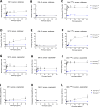
Fig 1
Fig 1. The fibrin(ogen) AGDV binding motif is required for both S. aureus adhesion to immobilized fibrinogen and clumping in fibrinogen solution.
Adhesion of stationary phase (A) WT S. aureus USA300, (B) ClfA- S. aureus USA300, and (C) ClfA-pclfa S. aureus USA300 to immobilized fibrinogenγWT or fibrinogenγΔ5. Clumping of stationary phase (D) WT S. aureus USA300, (E) ClfA- S. aureus USA300, or (F) ClfA-pclfa S. aureus USA300 in solutions containing fibrinogenγWT or fibrinogenγΔ5. Adhesion of exponential phase (G) WT S. aureus USA300, (H) to ClfA- S. aureus USA300, or (I) ClfA-pclfa S. aureus USA300 to immobilized fibrinogenγWT or fibrinogenγΔ5. Clumping of exponential phase (J) WT S. aureus USA300, (K) ClfA- S. aureus USA300, or (L) ClfA-pclfa S. aureus USA300 in solutions containing fibrinogenγWT or fibrinogenγΔ5. Data is derived from N = 3 replicates per fibrinogen concentration per group and presented as mean absorbance (for adhesion experiments) or the inverse of mean absorbance (for clumping experiments) ± SEM. Statistical significance was determined by 2- way ANOVA with Šídák’s multiple comparisons test where + = <0.05, * = <0.01, ^ = <0.001, and # = <0.0001.
Fig 2
Fig 2. FibγΔ5 mice display decreased survival following S. aureus peritonitis.
(A) Kaplan-Meyer log-rank analysis of FibγWT (n = 20) and FibγΔ5 (n = 20) mice following i.p. infection with 4.7x10⁸ CFUs of S. aureus USA300. (B) Evans Blue vascular leak assay on FibγWT and FibγΔ5 mice following i.p. injection with PBS or ~1x10⁹ CFU of S. aureus USA300. Fibrinogen ELISA on lavage fluid (C) and plasma (D) following 1 hour i.p. infection with S. aureus USA300. Data are presented as mean ± SEM and statistical significance was determined by 2-way ANOVA with Šídák’s multiple comparisons test.
Fig 3
Fig 3. FibγΔ5 mice display increased S. aureus CFUs in the peritoneal cavity following infection.
(A) Total bacterial CFUs in peritoneal lavage fluid of FibγWT and Fibγ Δ5 mice 1 hour after i.p. infection with 5x10⁸ CFUs of WT USA300 S. aureus. (B) Representative images of cytospin preparations of peritoneal lavage fluid collected from FibγWT and Fibγ Δ5 mice 1hr after i.p. infection with 5x10⁸ CFUs USA300 S. aureus. Note the presence of extensive free-floating bacteria in Fibγ Δ5 mice compared to FibγWT. (C) Total CFU in the peritoneal lavage fluid of FibγWT and FibγΔ5 mice 4 hours after i.p. infection with 4x10⁸ CFUs of WT USA300 S. aureus. (D) Representative images of cytospin preparations of peritoneal lavage fluid collected from FibγWT and FibγΔ5 mice 4 hours after infection. CFU data is presented as mean ± SEM and statistical significance was determined by Mann-Whitney u-test. Dashed horizontal lines indicate the infection dose. Images were captured with 40x objective with scale bar representing 20 μm. Total cell counts from peritoneal lavage fluid 1 hour (E) and 4 hours (F) after infection with USA300 S. aureus. Data are presented as mean ± SEM and statistical significance was determined by Mann-Whitney u-test. Differential cell counts from peritoneal lavage fluid 1 hour (G) and 4 hours (H) after infection. Data are presented as mean ± SEM and statistical significance was determined by 2-way ANOVA with Šídák’s multiple comparisons test. Analysis of total cells with internalized bacteria 1 hour (I) and 4 hours (J) after infection. Data are presented as mean ± SEM and statistical significance was determined by Mann-Whitney u-test.
Fig 4
Fig 4. FibγΔ5 mice display increased S. aureus dissemination to organ tissues.
Total CFUs in (A) blood, (B) heart and (C) lung of FibγWT and FibγΔ5 mice 1 hour after i.p. infection with 5x10⁸ CFUs of WT S. aureus USA300. Total CFUs in (D) blood, (E) heart and (F) lung of FibγWT and FibγΔ5 mice 4 hours after i.p. infection with 4x10⁸ CFUs of WT S. aureus USA300. Data are represented as mean ± SEM and statistical significance was determined by Mann-Whitney u-test.
Fig 5
Fig 5. Platelets but not platelet-derived protease-activated receptor-4 are important for local S. aureus clearance from the peritoneal cavity.
(A) Flow-cytometry analysis of platelet depletion following i.v. injection with anti-GPIb-α antibodies. Total bacterial CFUs in the peritoneal (B) lavage fluid, (C) blood, (D) heart and (E) lung of sham injected and platelet-depleted mice 1 hour after i.p. infection with 8x10⁸ CFUs of WT S. aureus USA300. (F) Total cell counts from peritoneal lavage fluid of sham injected and platelet-depleted mice 1 hour after infection with WT S. aureus USA300. Total bacteria CFUs in the peritoneal lavage (G) fluid, (H) blood, (I) heart and (J) lung of PAR4fl/fl and Pf4-cre+/PAR4fl/fl mice 1hr after i.p. infection with 6.57x10⁸ CFUs of WT S. aureus USA300. (K) Total cell counts from peritoneal lavage fluid of PAR4fl/fl and Pf4-Cre+/PAR4fl/fl mice 1 hour after infection with WT S. aureus USA300. Dashed horizontal lines indicate the infection dose. Data are presented as mean ± SEM and statistical significance was determined by Mann-Whitney u-test.
Fig 6
Fig 6. Deletion of ClfA from S. aureus USA300 results in increased S. aureus CFUs in the peritoneal cavity and a transient increase in CFUs the bloodstream and distant organs.
Total live bacteria in (A) lavage, (B) blood, (C) heart and (D) Lung of WT mice 1 hour after i.p. infection with WT (1x10⁹ CFUs) or ClfA- (7.8x10⁸ CFUs) S. aureus USA300. (E) Total host cells, (F) host cell differentials, and (G) and percentage of host cells with internalized bacteria in lavage fluid at 1 hour after infection. Total live bacteria in (H) lavage, (I) blood, (J) heart and (K) lung of WT mice 4 hours after i.p. infection with WT (1.6x10⁹ CFUs) or ClfA- (1.2x10⁹ CFUs) S. aureus USA300. (L) Total host cells, (M) host cell differentials, and (N) and percentage of host cells with internalized bacteria in lavage fluid at 1 hour after infection. CFU data are presented as mean ± SEM and statistical significance was determined by Mann-Whitney u-test. Host cell counts and percentages are presented as mean ± SEM with statistical significance determined by 2-way ANOVA with Šídák’s multiple comparisons test. Data on cells with internalized bacteria are presented as mean ± SEM and statistical significance was determined by Mann-Whitney U-test. Dashed horizontal lines indicate the infection dose.
Fig 7
Fig 7. Fibrin-macrophage driven host antimicrobial function limits dissemination, but fibrin matrix formation alone is not sufficient to suppress systemic spread of S. aureus out of the peritoneal cavity.
Total live bacteria in (A) lavage fluid, (B) blood, (C) heart and (D) lung and total host cell counts (E) from WT and FibAEK mice 1 hr after infection with 8.8x10⁸ CFUs of WT S. aureus USA300. Total live bacteria in lavage fluid (F), blood (G), heart (H) and lung (I) and total host cell counts (J) from WT and FXIII-/- mice 1 hr after infection with 1x10⁹ CFUs of WT S. aureus USA300. Total live bacteria in lavage fluid (K), blood (L), heart (M) lung (N) and total host cell count (O) from WT mice treated with PBS or Clodronate liposomes 1 hr after infection with 9x10⁸ CFUs of WT S. aureus USA300. Total live bacteria in lavage fluid (P), blood (Q), heart (R) and lung (S) and total host cell counts (T) from WT and Fibγ390-396A mice 1 hr after infection with 6.7x10⁸ CFUs of WT S. aureus USA300. Data are presented as mean ± SEM and statistical significance was determined by Mann-Whitney u-test.

Similar articles

Cited by

References

    1. Brown AF, Leech JM, Rogers TR, McLoughlin RM. Staphylococcus aureus Colonization: Modulation of Host Immune Response and Impact on Human Vaccine Design. Front Immunol. 2014;4:507. doi: 10.3389/fimmu.2013.00507 - DOI - PMC - PubMed
    1. Lowy FD. Staphylococcus aureus infections. N Engl J Med. 1998;339(8):520–32. doi: 10.1056/NEJM199808203390806 - DOI - PubMed
    1. Tong SY, Davis JS, Eichenberger E, Holland TL, Fowler VG Jr. Staphylococcus aureus infections: epidemiology, pathophysiology, clinical manifestations, and management. Clin Microbiol Rev. 2015;28(3):603–61. doi: 10.1128/CMR.00134-14 - DOI - PMC - PubMed
    1. Melzer M, Welch C. Thirty-day mortality in UK patients with community-onset and hospital-acquired meticillin-susceptible Staphylococcus aureus bacteraemia. J Hosp Infect. 2013;84(2):143–50. doi: 10.1016/j.jhin.2012.12.013 - DOI - PubMed
    1. Pottinger PS. Methicillin-resistant Staphylococcus aureus infections. Med Clin North Am. 2013;97(4):601–19, x. doi: 10.1016/j.mcna.2013.02.005 - DOI - PubMed

Publication types