. 2022 Jan;12(1):210264.
doi: 10.1098/rsob.210264. Epub 2022 Jan 19.

Mapping of a N-terminal α-helix domain required for human PINK1 stabilization, Serine228 autophosphorylation and activation in cells

Affiliations

Mapping of a N-terminal α-helix domain required for human PINK1 stabilization, Serine228 autophosphorylation and activation in cells

Poonam Kakade et al. Open Biol. 2022 Jan.

Abstract

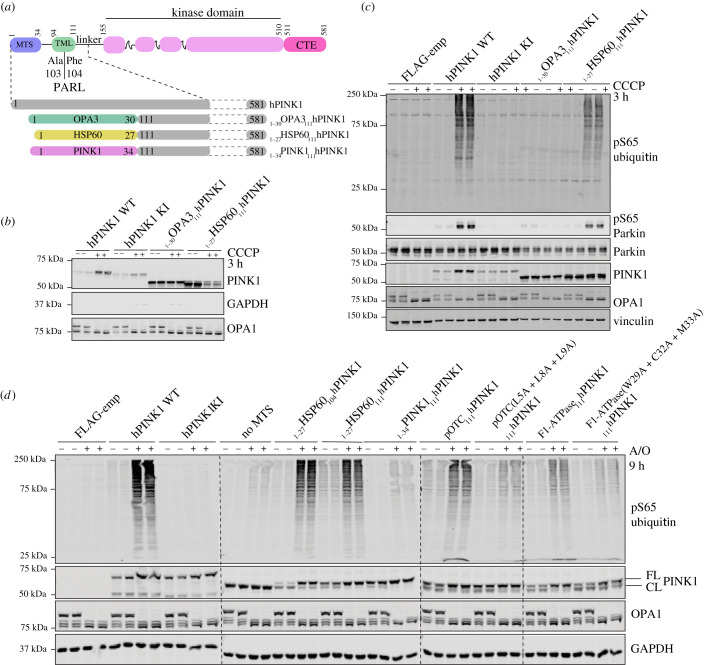
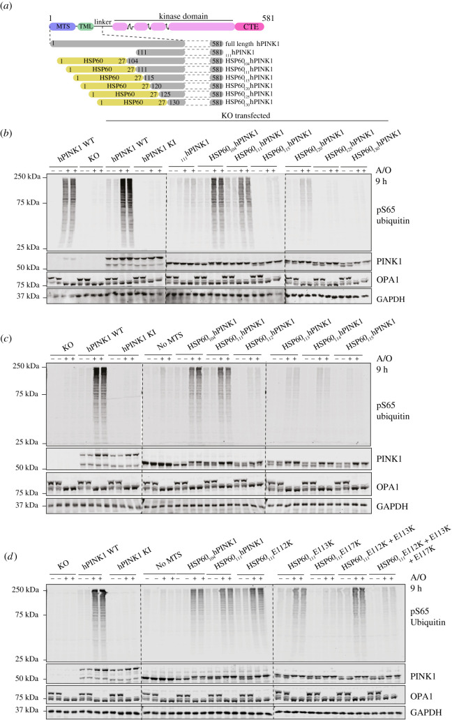
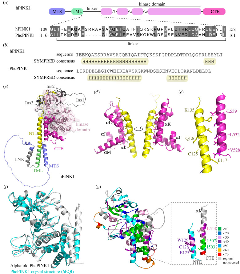
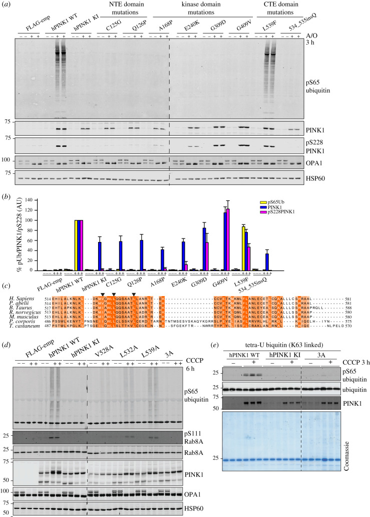
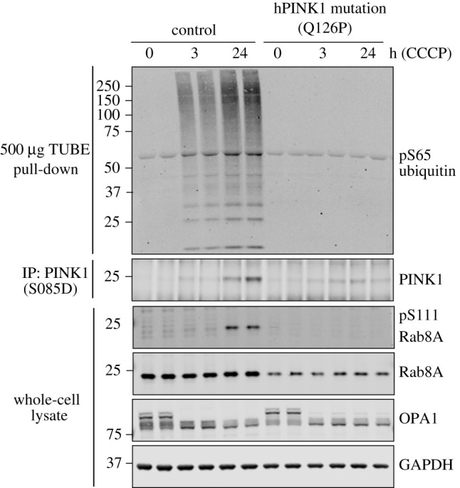
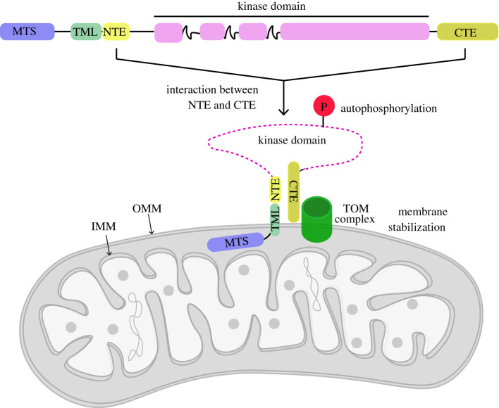
Autosomal recessive mutations in the PINK1 gene are causal for Parkinson's disease (PD). PINK1 encodes a mitochondrial localized protein kinase that is a master-regulator of mitochondrial quality control pathways. Structural studies to date have elaborated the mechanism of how mutations located within the kinase domain disrupt PINK1 function; however, the molecular mechanism of PINK1 mutations located upstream and downstream of the kinase domain is unknown. We have employed mutagenesis studies to define the minimal region of human PINK1 required for optimal ubiquitin phosphorylation, beginning at residue Ile111. Inspection of the AlphaFold human PINK1 structure model predicts a conserved N-terminal α-helical extension (NTE) domain forming an intramolecular interaction with the C-terminal extension (CTE), which we corroborate using hydrogen/deuterium exchange mass spectrometry of recombinant insect PINK1 protein. Cell-based analysis of human PINK1 reveals that PD-associated mutations (e.g. Q126P), located within the NTE : CTE interface, markedly inhibit stabilization of PINK1; autophosphorylation at Serine228 (Ser228) and Ubiquitin Serine65 (Ser65) phosphorylation. Furthermore, we provide evidence that NTE and CTE domain mutants disrupt PINK1 stabilization at the mitochondrial Translocase of outer membrane complex. The clinical relevance of our findings is supported by the demonstration of defective stabilization and activation of endogenous PINK1 in human fibroblasts of a patient with early-onset PD due to homozygous PINK1 Q126P mutations. Overall, we define a functional role of the NTE : CTE interface towards PINK1 stabilization and activation and show that loss of NTE : CTE interactions is a major mechanism of PINK1-associated mutations linked to PD.

Keywords: PINK1; Parkinson's disease; kinase; mitochondria; phosphorylation; translocase.


Conflict of interest statement

M.M.K.M. is a member of the Scientific Advisory Board of Mitokinin Inc.

Figures

Figure 1.
Figure 1.
MTS that mediate TOM complex association are required for PINK1 activation. (a) Schematic depiction of constructs for human PINK1 (hPINK1) mutants. For hPINK1 mutants targeted to different compartments of the mitochondria, residues 1–110, which includes the MTS and transmembrane-like domain (TML) and PARL cleavage site of hPINK1, were removed and replaced with the predicted MTS of proteins known to localize to the OMM (OPA3 1–30) and matrix (HSP60 1–27). For hPINK1 mutant with predicted MTS of PINK1, residues 1–110 were replaced with hPINK 1 residues 1–34. (b) Flp-In TRex HEK293 hPINK1 KO cells stably expressing WT, KI (D384A), OPA3 (1–30)-hPINK1(111–581) or HSP60 (1–27) hPINK1(111–581) were treated ±10 µM CCCP for 3 h and subjected to sub-cellular fractionation to obtain membrane-enriched fraction. Samples were resolved by SDS-PAGE. Proteins were transferred to nitrocellulose membranes probed using the antibodies indicated. n = 2. (c) Flp-In TRex HEK293 cells PINK1 KO stably expressing empty vector (FLAG-emp), hPINK1-3FLAG WT, kinase inactive (KI, D384A), OPA3 (1–30)-hPINK1 (111–581) and HSP60 (1–27)-hPINK1 (111–581) were treated with ±10 µM CCCP for 3 h. Whole-cell lysates (Parkin and control blots) or membrane-enriched fractions (FLAG and pSer65 ubiquitin blots) were resolved by SDS-PAGE. Proteins were transferred to nitrocellulose membranes probed using the antibodies indicated. (d) Human SK-OV-3 PINK1 knockout cells were transfected with empty vector (FLAG-emp), hPINK1-3FLAG WTkinase inactive (KI, D384A), HSP60 (1–27)-hPINK1 (104–581), HSP60 (1–27)-hPINK1 (111–581), hPINK1 (1–34)-hPINK1 (111–581), pOTC (1–30)-hPINK1 (111–581) and pF1ATPase (1–30)-hPINK1 (111–581). Cells were treated with A/O for 9 h prior to lysis and probed using the indicated antibodies.
Figure 2.
Figure 2.
Mapping of N-terminal boundary of minimal region required for PINK1 activation to residue Ile111. (a) Schematic depiction of deletion mutants of human PINK1 (hPINK1) (start sites varying from 104–130 to END) fused at the N-terminus with HSP60-MTS aa 1–27. (b) Cell-based analysis of WT and hPINK1 knockout (PINK1 KO) SK-OV-3 cells transfected with indicated deletion mutants of hPINK1 ranging from HSP60(1–27)-hPINK1(104–581) to HSP60(1–27)- hPINK1(130–581) alongside full-length WT and kinase inactive (KI) hPINK1. Cells were treated with A/O for 9 h prior to lysis and immunoblotted with indicated antibodies (anti-pSer65 ubiquitin, anti-PINK1, anti-OPA1 and anti-GAPDH). The membranes were developed using the LI-COR Odyssey CLx Western blot imaging system. (c) Cell-based analysis of PINK1 KO SK-OV-3 cells transfected with deletion mutants of hPINK1 ranging from HSP60(1–27)-hPINK1(111–581) to HSP60(1–27)-hPINK1(115–581). Cells were treated with A/O for 9 h prior to lysis and immunoblotting with indicated antibodies and analysed as described above. (d) Cell-based analysis of PINK1 KO SKOV3 cells transfected with N-terminal hPINK1 E-K single mutants (E112K or E113K or E117K), double mutant (E112K + E113K) and triple mutants (E112K + E113K + E117K) of the region spanning residues 111–117 of HSP60(1–27)-hPINK1(111–581). Cells were treated with A/O for 9 h prior to lysis, blotted with indicated antibodies and analysed as described above.
Figure 3.
Figure 3.
Structural modelling predicts an N-terminal α-helix extension (NTE domain) and its interaction with CTE domain. (a) Prediction of α-helix in the linker region of PINK1 flanked by transmembrane domain and kinase domain. Schematic representation of human PINK1 (hPINK1) and Pediculus humanus corporis PINK1 (PhcPINK1) highlighting the linker region flanked by transmembrane domain and kinase domain. The alignment of linker region is performed by MAFT and annotated in Jalview. (b) Prediction of secondary structure in linker region of hPINK1 and PhcPINK1 by SYMPRED. SYMPRED predicted α-helix represents a consensus prediction of α-helix by different prediction tools; PROF, SSPRO, YASPIN, JNET and PSIPRED. Predicted α-helices are highlighted by cylinders. (c) Complete structure of human PINK1 (hPINK1) solved by AlphaFold (Uniprot ID: Q9BXM7) and colour coded according to regions. (e) α-NTE domain of hPINK1 forms intramolecular interaction with CTE domain. α-NTE region primarily interacts with αK region of CTE. (e) Location of conserved residues at the NTE:CTE interface; PD-associated residues are highlighted. The residues of α-NTE at the start of interface (E117) and end of α-helix (K135) are also highlighted. (f) AlphaFold predicted PhcPINK1(108–575) model (grey) superposed with the crystal structure of PhcPINK1 (6EQI, blue) with a rmsd of 0.7. The missing NTE region in the crystal structure is labelled. (g) HDX data mapped on AlphaFold modelled PhcPINK1(108–575) with zoomed in view of NTE and CTE. The percentage deuterium uptake across the protein after 2 min of labelling is colour-coded as labelled. Residues also colour-coded according to the percentage deuterium uptake.
Figure 4.
Figure 4.
Mutational analysis confirms the critical role of NTE and CTE domains for PINK1 activation. (a) α-NTE and CTE PD-associated mutants lead to reduced protein stabilization, and loss of autophosphorylation and hPINK1 activation. Stably expressing PINK1-3FLAG WT, KI (D384A), empty vector (FLAG-emp), α-NTE mutants (C125G, Q126P), kinase domain mutants (A168P, E240K, G309D, G409V) and CTE domain mutants (L539F, ins534Q) cell lines were generated in PINK1-knockout Flp-In TRex-HeLa cells. PINK1-3FLAG expression was induced by 24 h treatment with 0.2 uM doxycycline, and mitochondrial depolarization induced by 3 h treatment with 10 µM A/O where indicated. Mitochondrial-enriched fractions were subjected to immunoblotting with α-PINK1 (in-house/DCP antibody), α-ubiquitin pS65 (CST), α-OPA1 (BD) and α-HSP-60 primary antibodies. n = 3. (b) Immunoblots were quantified for phospho-Ser65 Ub/HSP-60, PINK1/HSP-60 and pS228/HSP-60 using Image Studio software. Data are presented relative to WT hPINK as mean ± s.d. (n = 3). (c) Multiple sequence alignment of CTE region of PINK1 orthologues across species. Sequence alignment was performed with MUSCLE and annotated in Jalview. Mutated CTE residues for functional analysis are highlighted with arrow heads. (d) CTE mutants exhibit reduced stabilization, autophosphorylation and substrate phosphorylation. hPINK1 knockout HeLa cells transiently expressing hPINK1-3FLAG WT, KI or hPINK1 CTE mutants V528A (L503A in PhcPINK1), L532A (L507A in PhcPINK1) and L539A (L514A in PhcPINK1). Cells were stimulated ±10 μM CCCP for 6 h. Membrane fractions were isolated and solubilized in 1% Triton X-100 lysis buffer Lysates that were resolved by SDS-PAGE. Proteins were transferred to nitrocellulose membranes probed using the antibodies indicated. n = 2. (e) CTE triple mutant is inactive against recombinant substrates in vitro. Flp-In HEK293 PINK1 KO cells stably expressing WT or KI (D384A) or dimerization triple mutant (V528A/L532A/L539A) PINK1 were treated ±10 µM CCCP for 3 h and subjected to sub-cellular fraction in a CFAB. Three micrograms of non-solubilized membrane-enriched fraction (MeF) were incubated with tetraubiquitin (K63 linked) in CFAB supplemented with 2 mM (unlabelled) ATP, 5 mM MgCl2, 2 mM DTT and 0.75% Glycerol for 20 min. Samples were resolved via SDS-PAGE and blotted for the indicated antibodies. n = 2.
Figure 5.
Figure 5.
Endogenous PINK1 stabilization and activation is reduced in Q126P PINK1 patient-derived fibroblasts. Primary fibroblast cultures established from skin biopsies from a healthy subject (control) and a PD patient harbouring a PINK1 Q126P homozygous mutation were treated with 10 μM CCCP or DMSO for 3 h or 24 h. Lysates were subjected to immunoblot analysis using the indicated antibodies. Ubiquitin capture was used prior to immunoblotting with anti-phospho-Ser65 ubiquitin antibody. Endogenous PINK1 was detected after immunoprecipitation from whole-cell lysate.
Figure 6.
Figure 6.
Schematic of role of NTE : CTE interaction towards PINK1 stabilization at mitochondria. NTE domain interaction with CTE facilitates stabilization and activation of PINK1 at TOM complex. Abbreviations: mitochondrial-targeting sequence (MTS); transmembrane-like domain (TML); N-terminal extension domain (NTE); C-terminal extension (CTE); translocase of outer membrane (TOM); inner mitochondrial membrane (IMM); outer mitochondrial membrane (OMM).

References

    1. Valente EM, et al. 2004. Hereditary early-onset Parkinson's disease caused by mutations in PINK1. Science 304, 1158-1160. (10.1126/science.1096284) - DOI - PubMed
    1. Woodroof HI, et al. 2011. Discovery of catalytically active orthologues of the Parkinson's disease kinase PINK1: analysis of substrate specificity and impact of mutations. Open Biol. 1, 110012. (10.1098/rsob.110012) - DOI - PMC - PubMed
    1. Yamano K, Youle RJ. 2013. PINK1 is degraded through the N-end rule pathway. Autophagy 9, 1758-1769. (10.4161/auto.24633) - DOI - PMC - PubMed
    1. Kondapalli C, et al. 2012. PINK1 is activated by mitochondrial membrane potential depolarization and stimulates Parkin E3 ligase activity by phosphorylating Serine 65. Open Biol. 2, 120080. (10.1098/rsob.120080) - DOI - PMC - PubMed
    1. Harper JW, Ordureau A, Heo JM. 2018. Building and decoding ubiquitin chains for mitophagy. Nat. Rev. Mol. Cell Biol. 19, 93-108. (10.1038/nrm.2017.129) - DOI - PubMed

Publication types