. 2022 Jan 18;18(1):44.
doi: 10.1186/s12917-021-03091-6.

Osteochondrosis and other lesions in all intervertebral, articular process and rib joints from occiput to sacrum in pigs with poor back conformation, and relationship to juvenile kyphosis

Affiliations

Osteochondrosis and other lesions in all intervertebral, articular process and rib joints from occiput to sacrum in pigs with poor back conformation, and relationship to juvenile kyphosis

Kristin Olstad et al. BMC Vet Res. .

Abstract

Background: Computed tomography (CT) is used to evaluate body composition and limb osteochondrosis in selection of breeding boars. Pigs also develop heritably predisposed abnormal curvature of the spine including juvenile kyphosis. It has been suggested that osteochondrosis-like changes cause vertebral wedging and kyphosis, both of which are identifiable by CT. The aim of the current study was to examine the spine from occiput to sacrum to map changes and evaluate relationships, especially whether osteochondrosis caused juvenile kyphosis, in which case CT could be used in selection against it. Whole-body CT scans were collected retrospectively from 37 Landrace or Duroc boars with poor back conformation scores. Spine curvature and vertebral shape were evaluated, and all inter-vertebral, articular process and rib joints from the occiput to the sacrum were assessed for osteochondrosis and other lesions.

Results: Twenty-seven of the 37 (73%) pigs had normal spine curvature, whereas 10/37 (27%) pigs had abnormal curvature and all of them had wedge vertebrae. The 37 pigs had 875 focal lesions in articular process and rib joints, 98.5% of which represented stages of osteochondrosis. Five of the 37 pigs had focal lesions in other parts of vertebrae, mainly consisting of vertebral body osteochondrosis. The 10 pigs with abnormal curvature had 21 wedge vertebrae, comprising 10 vertebrae without focal lesions, six ventral wedge vertebrae with ventral osteochondrosis lesions and five dorsal wedge vertebrae with lesions in the neuro-central synchondrosis, articular process or rib joints.

Conclusions: Computed tomography was suited for identification of wedge vertebrae, and kyphosis was due to ventral wedge vertebrae compatible with heritably predisposed vertebral body osteochondrosis. Articular process and rib joint osteochondrosis may represent incidental findings in wedge vertebrae. The role of the neuro-central synchondrosis in the pathogenesis of vertebral wedging warrants further investigation.

Keywords: Helical computed tomography; Juvenile (Scheuermann’s) kyphosis; Neuro-central synchondrosis; Osteochondrosis; Swine; Vascular failure; Wedge vertebra; Zygapophyseal joint.


Conflict of interest statement

The authors declare that they have no competing interests.

Figures
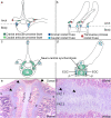
Fig. 1
Fig. 1
Vertebral anatomy, ossification centres and growth cartilage. a-b. Vertebrae consist of a body and a neural arch. b. The cranial and caudal articular facets (dark and light green) meet at the articular process joints. b. Adjacent vertebral bodies articulate with the head of a rib at the caudal and cranial costal foveae (dark and light blue) to form the costo-vertebral joint. a-b. The rib tubercle articulates with the costal fovea of the transverse process (red) to form the costo-transverse joint. c-d. The primary ossification centre (1°) is responsible for forming the ventral 2/3 of the vertebral midbody, whereas d. the secondary ossification centres (2°) cover the entire cranial and caudal ends of the vertebral body. c-d. The body and arch meet at the neuro-central synchondrosis (labelled). c. The neural arch forms from a pair of primary ossification centres (1°) located within the left and right demi-arches, respectively, and c-d. the spinous process, transverse process and cranial and caudal articular processes have secondary ossification centres (2°). d-e. Between primary and secondary ossification centres, there are metaphyseal growth plates or physes (P) and superficial to secondary ossification centres, there is epiphyseal growth cartilage (EGC). e-f. The growth cartilage of the physes and synchondrosis (S) contains patent vessels or eosinophilic streaks (arrowheads) representing the cartilage canal blood supply. f. The synchondrosis is bidirectional and contains one proliferative zone (PFZ1) contributing to the neural arch and a second proliferative zone (PFZ2) forming the dorsal 1/3 of the vertebral midbody. a-b. Tracing of T7 from photograph; a. cranial view, b. lateral view. c-d. Simplified indication of ossification centres from CT scans on top of tracings; a. mid-body transverse plane, b. lateral view. e-f. Para-sagittal histological section from T4-T5 of a 12 kg mixed-breed piglet; e. 10x; f. 100x magnification, haematoxylin and eosin
Fig. 2
Fig. 2
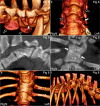
Transitional vertebrae with characteristics of both the cranially and caudally adjacent segments. a. Pig 14 has a transitional vertebra T15 at the thoraco-lumbar junction with both a left and a right rib (arrows), and five lumbar vertebrae. The spine curvature is normal.b. Pig 4 has a transitional vertebra T16 at the thoraco-lumbar junction that is asymmetric in the sense that it only has one rib on the left side (arrow), but spine curvature is normal. a-b. 3D model, dorsal view
Fig. 3
Fig. 3
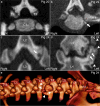
Articular process and rib joint osteochondrosis, osteochondrosis dissecans (OCD), cyst and osteoarthritis. a. Pig 20. There is a multi-focal, sharply demarcated, uniformly hypodense defect (dashed lines) categorised as a multi-lobulated osteochondrosis lesion in the right caudal articular process of C7. b. Pig 22. There is a mineral hyperdense body (arrow) categorised as an OCD lesion in the joint between the caudal costal facet of T3 and the head of the fourth rib on the left side. c. Pig 24. There is a roughly spherical defect (arrow) categorised as a cyst in the left caudal articular process of L2. d-e. Pig 24. The joint space is irregular, and there is subchondral bone sclerosis (arrows) and periarticular osteophytes (arrowheads) categorised as osteoarthritis of the left articular process joint of L3-L4. a-b. Transverse plane. c-d. Dorsal plane. e. 3D model, left and slightly left-dorsal-to-right-ventral oblique view
Fig. 4
Fig. 4
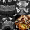
Osteochondrosis dissecans (OCD) in the neuro-central synchondrosis of pig 5. a. There is an OCD lesion (arrow) located towards the floor of the vertebral canal caudally in C6. a. The lesion (arrow) is located cranial to the caudal physis of C6, and b-d. near the midline, corresponding to the neuro-central synchondrosis. a. Sagittal plane. b. Dorsal plane. c. Transverse plane. d. 3D model, caudal and slightly right-to-left oblique view
Fig. 5
Fig. 5
Pathological Type I Salter-Harris fracture of the neuro-central synchondrosis of pig 7*. a. The neuro-central synchondroses (arrows) caudally in T7 were 2–3 times normal thickness. b. Image of the neuro-central synchondroses (arrows) of T8 for comparison. c. The increased thickness (arrow) was interpreted as a pathological, Type I Salter-Harris fracture of the neuro-central synchondrosis in T7 secondary to the visible ventral wedging of T6 and dorsal wedging of T8. d. The wedging is associated with marked kyphosis and lordosis. a-b. Transverse plane. c. Sagittal plane. c. 3D model, left view
Fig. 6
Fig. 6
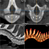
Ventral vertebral body osteochondrosis and kyphosis in pig 11. a. There is an osteochondrosis lesion (arrow) affecting both the physis and the epiphysis caudo-ventrally in T16. b. The osteochondrosis lesion (arrow) is located to the right of the midline. c. The lesion (dashed lines) is located between 5 and 8 o’clock on a clockface (circle and 3, 6, 9 and 12 indicators) superimposed on the vertebral body in the transverse plane. d. There is mild kyphosis centred at T16 with the osteochondrosis lesion (arrow). a. Sagittal plane. b. Dorsal plane. c. Transverse plane. d. 3D model, left and slightly left-ventral-to-right-dorsal oblique view
Fig. 7
Fig. 7
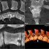
Multiple vertebral body osteochondrosis lesions, osteochondrosis dissecans (OCD), cyst, spondylosis, wedging and kyphosis in pig 21. a. There are osteochondrosis lesions (arrows) cranio- and caudo-ventrally in T14, an OCD lesion (arrowhead) cranio-ventrally in T13 and a cyst (open arrow; for non-tangential section, see c.) caudally in T15, all of which are also ventral wedge vertebrae. A smooth, bony spur (open arrowhead) extends from the cranio-ventral aspect of T14 towards the caudo-ventral aspect of T13, interpreted as bone bridge formation/spondylosis. b. The osteochondrosis lesion (arrows) cranially in T14 is multi-lobulated and located to the right of the midline. The mineral opacity (arrowhead) superficial to the lesion is cut tangentially here and in a. and was connected to the parent bone in other planes of section, interpreted as reparative ossification. c. Para-sagittal slice through the centre of the cyst (arrows) caudally in T15. d. There is mild kyphosis centred at the T14-T15 junction (arrow). a-c. Sagittal plane. b. Dorsal plane. d. 3D model, left view
Fig. 8
Fig. 8
Cervical transverse process “transposition”, block vertebra and kyphosis in pig 8. a-b. The left transverse process of C6 (arrow) is small and appears to be fused (arrowhead) to the transverse process of C7. The left transverse process of C5 (open arrow) is large and matches the right transverse process of C6 (open arrowhead) in size, compatible with malformation/“transposition” of the left transverse process of C6 to C5. c. The intervertebral disc and secondary ossification centres are absent between the vertebral bodies of T13–14, which are fused into a ventrally wedged block vertebra. d. An oblique, hyperdense line (between arrows) marks the junction between the primary ossification centres, and T14 is located slightly to the right of T13, d. e. causing the spinal axis to shift to the right from T14 caudally. e. The block vertebra articulates with rib pairs 13, 14 and 15. f. There is marked kyphosis centred at the T13–14 block vertebra. a, f. 3D models, left views. b, e. 3D models, ventral views. c. Sagittal plane. d. Dorsal plane

References

    1. Nielsen LW, Hogedal P, Arnbjerg J, Jensen HE. Juvenile kyphosis in pigs. A spontaneous model of Scheuermann's kyphosis. APMIS. 2005;113(10):702–707. - PubMed
    1. Lahrmann KH, Hartung K. Causes of kyphotic and lordotic curvature of the spine with cuneiform vertebral deformation in swine. Berl Munch Tierarztl Wochenschr. 1993;106(4):127–132. - PubMed
    1. Grøndalen T. Osteochondrosis and arthrosis in pigs. I. Incidence in animals up to 120 kg live weight. Acta Vet Scand. 1974;15(1):1–25. - PMC - PubMed
    1. Straw B, Bates R, May G. Anartomical abnormalities in a group of finishing pigs: prevalence and pig performance. J Swine Health Prod. 2009;17(1):28–31.
    1. Corradi A, Alborali L, Passeri B, Salvini F, De Angelis E, Martelli P, et al. Acquired hemivertebrae in «humpy-backed» piglets. In: 18th IPVS Congress: 2004. Hamburg: International Pig Veterinary Society; 2004. p. 357.

LinkOut - more resources