. 2022 Jan 19;20(1):29.
doi: 10.1186/s12916-022-02240-4.

Single-dose SARS-CoV-2 vaccinations with either BNT162b2 or AZD1222 induce disparate Th1 responses and IgA production

Affiliations

Single-dose SARS-CoV-2 vaccinations with either BNT162b2 or AZD1222 induce disparate Th1 responses and IgA production

Michael Müller et al. BMC Med. .

Abstract

Background: While vaccination programs against the severe acute respiratory syndrome virus 2 (SARS-CoV-2) are globally ongoing, disparate strategies for the deployment of spike antigen show varying effectiveness.

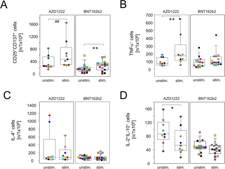
Methods: In order to explore this phenomenon, we sought to compare the early immune responses against AZD1222 and BNT162b2. SARS-CoV-2 seronegative participants received a single dose of either vaccine and were analyzed for immune cell, effector T cell, and antibody dynamics.

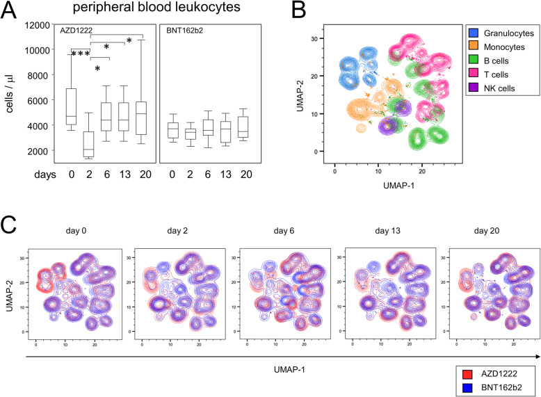
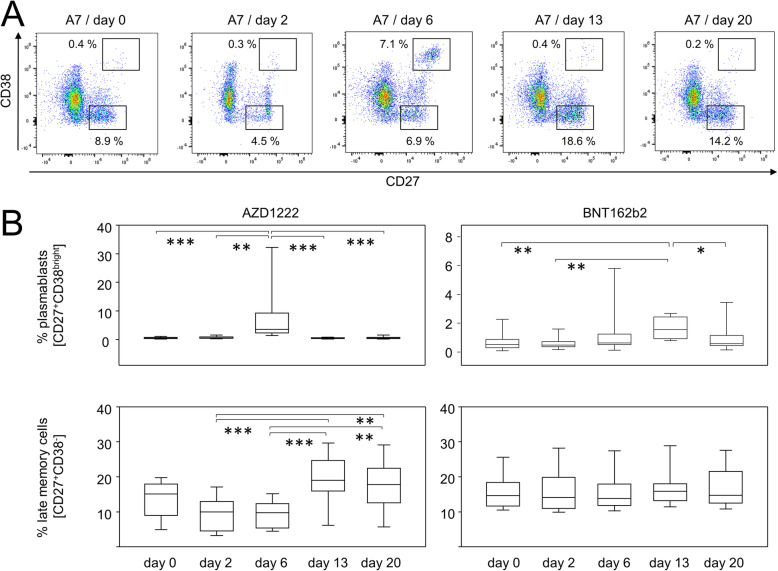
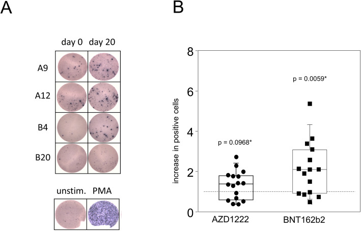
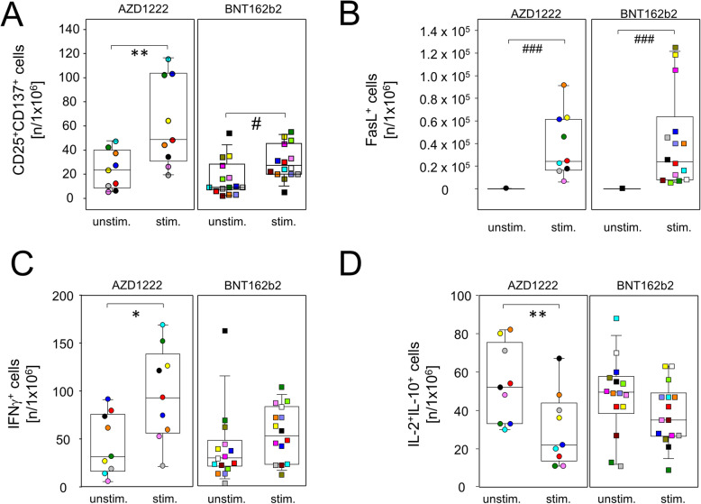
Results: AZD1222 induced transient leukopenia and major changes among innate and adaptive subpopulations. Both vaccines induced spike protein-specific effector T cells which were dominated by type 1 helper T cell responses following AZD1222 vaccination. A significant reduction of anti-inflammatory T cells upon re-stimulation was also restricted to AZD1222 vaccinees. While IgM and IgG were the dominant isotypes elicited by AZD1222, BNT162b2 led to a significant production of IgG and IgA.

Conclusions: Our results suggest that the strategy for spike protein delivery impacts on how and to what extent immune priming against the main SARS-CoV-2 antigen proceeds.

Keywords: AZD1222; BNT162b2; COVID-19; Cytotoxic T cells; SARS-CoV-2; Type 1 helper T cells; Vaccination.


Conflict of interest statement

The authors declare that they have no competing interests.

Figures

Fig. 1
Fig. 1
Vaccination with AZD1222 induced a transient reduction of peripheral leukocytes and displacements of major immune cell populations. A Leukocyte counts in the peripheral blood after vaccination with AZD1222 (n = 13, left panel) and BNT162b2 (n = 15, right panel). p-values resulting from multiple group comparisons were 0.0005 (AZD1222/Kruskal-Wallis and Dunn’s multiple comparisons tests) and 0.5306 (BNT162b2/one-way ANOVA), respectively. **p < 0.01, ***p < 0.001. B UMAP of surface antigen expression and clustering of major immune cell populations for all time points after vaccination. C UMAPs on immune cell compositions for each time point
Fig. 2
Fig. 2
AZD1222 vaccination induced the enrichment of pro-inflammatory monocytes. A Pseudocolor plots for the expression of CD14 and CD16 on monocytes are representative for the AZD1222 vaccination group. B Proportions of CD16+CD14+ pro-inflammatory monocytes after vaccination with AZD1222 (n = 18, left panel) or BNT162b2 (n = 18, right panel). All FACS analyses were on gated live monocytes. Asterisks indicate significant differences between time points. p-values resulting from one-way ANOVA and Tukey-Kramer multiple comparisons tests were < 0.0001 for AZD1222 and 0.0146 for BNT162b2 analyses. ***p < 0.001
Fig. 3
Fig. 3
Vaccination with AZD1222 and BNT162b2 induced significant yet transient alterations among peripheral plasmablasts. AZD1222 also induced a significant increase in late memory B cells on days 13 and 20. A Pseudocolor plots for the expression of CD27 and CD38 on B cells are representative for the AZD1222 vaccination group. B Proportions of CD27+CD38bright plasmablasts (top) and CD27+CD38 late memory B cells (bottom) after vaccination with AZD1222 (n = 18, left) or BNT162b2 (n = 18, right). All FACS analyses were on CD19+CD45RA+ B cells. p-values resulting from one-way ANOVAs were < 0.0001 for both AZD1222 analyses. p-values resulting from Kruskal-Wallis and Dunn’s multiple comparisons tests were 0.0008 for the comparison of plasmablast and 0.6998 for the comparison of late memory B cells for the BNT162b2 analyses. *p < 0.05, **p < 0.01, ***p < 0.0001
Fig. 4
Fig. 4
Spike-specific IFNγ-producing lymphocytes were significantly increased after vaccination with BNT162b2. PBMCs were isolated at the day of vaccination (day 0) and 20 days later. Cells were then stimulated with recombinant SARS-CoV-2 spike protein for 24 h. Subsequently, an ELISPOT assay for the detection of IFNγ-producing cells was performed. A Representative shots of spot-forming cells from individuals vaccinated with either AZD1222 (A9 and A12) or BNT162b2 (B4 and B20). PBMCs were stimulated with PMA as a positive control (bottom). B The counts of spot-forming cells from day 20 were normalized to counts from day 0. Data show the relative increase in IFNγ+ cells after vaccination (n = 16 for AZD1222 and n = 15 for BNT162b2). p-values result from one-sample t-tests assessing the differences of group means to the hypothetical value of 1
Fig. 5
Fig. 5
Vaccination with single doses of AZD1222 or BNT162b2 induced the expansion of cytotoxic effector T cells. PBMCs were isolated on day 20 after vaccination and stimulated with (stim.) or without (unstim.) spike protein-encoding mRNA (BNT162b2) for 24 h and analyzed by flow cytometry for the expression of inducible activation markers and intracellularly trapped cytokines among CD8+ cells. Sample sizes were n = 9 for AZD1222 and n = 15 for BNT162b2, respectively. Paired samples are illustrated by color coding. In vitro re-stimulation significantly increased CD25+CD137+ (A) and FasL expressing cells in both vaccination groups (B). INFγ–producers were increased (C) and IL-2 and IL-10 co-expressing cells were significantly reduced among AZD1222 vaccinees only (D). *p < 0.05, **p < 0.01 resulting from paired t-tests. #p < 0.05, ###p < 0.001 resulting from Wilcoxon signed rank tests for matched pairs
Fig. 6
Fig. 6
AZD1222 vaccination supported the induction of type 1 helper T cells. PBMCs were isolated on day 20 after vaccination and stimulated with (stim.) or without (unstim.) spike protein encoding mRNA (BNT162b2) for 24 h and analyzed by flow cytometry for the expression of inducible activation markers and intracellularly trapped cytokines among CD4+ cells. Sample sizes were n = 9 for AZD1222 and n = 15 for BNT162b2, respectively. Paired samples are illustrated by color coding. In vitro re-stimulation significantly increased CD25+CD137+ cells in both vaccination groups (A) and TNFα-producing cells in the AZD1222 vaccinated group, only B. C Neither vaccination allowed for the re-stimulation of IL-4-producing cells. D IL-2 and IL-10 co-expressing cells were significantly reduced among AZD1222 vaccinees only. *p < 0.05, **p < 0.01, resulting from paired t-test. ##p < 0.01, resulting from Wilcoxon signed rank test for matched pairs
Fig. 7
Fig. 7
A single dose of BNT162b2 induced significantly higher amounts of spike protein binding IgG, IgA, and receptor binding domain neutralizing antibodies than AZD1222. A SARS-CoV-2 spike binding antibodies were detected by ELISA and absorbance readouts were normalized to calibrator values to obtain arbitrary units. Gray lines depict the positive response threshold within a range of 0.8 to 1.1 arbitrary units. Sample sizes were n = 18 for AZD1222 and n = 18 for BNT162b2, respectively. p-values resulting from one-way ANOVA and Tukey-Kramer multiple comparisons tests were < 0.0001 for all three isotype analyses. B Neutralizing antibodies were determined by the surrogate virus competitive ELISA. Absorbance readouts from BNT162b2 (n = 17) and AZD1222 (n = 17) were calibrated with WHO standards to obtain international units (IU)/mL. *p < 0.05 resulting from Mann-Whitney U test and ***p < 0.001 resulting from Dunn’s test

References

    1. Dong E, Du H, Gardner L. An interactive web-based dashboard to track COVID-19 in real time. Lancet Infect Dis. 2020;20(5):533–534. doi: 10.1016/S1473-3099(20)30120-1. - DOI - PMC - PubMed
    1. Muralidar S, Ambi SV, Sekaran S, Krishnan UM. The emergence of COVID-19 as a global pandemic: understanding the epidemiology, immune response and potential therapeutic targets of SARS-CoV-2. Biochimie. 2020;179:85–100. doi: 10.1016/j.biochi.2020.09.018. - DOI - PMC - PubMed
    1. Hanrath AT, Payne BAI, Duncan CJA. Prior SARS-CoV-2 infection is associated with protection against symptomatic reinfection. J Infect. 2021;82(4):e29–e30. doi: 10.1016/j.jinf.2020.12.023. - DOI - PMC - PubMed
    1. Okba NMA, Müller MA, Li W, Wang C, GeurtsvanKessel CH, Corman VM, et al. Severe acute respiratory syndrome coronavirus 2−specific antibody responses in coronavirus disease patients. Emerg Infect Dis. 2020;26(7):1478–1488. doi: 10.3201/eid2607.200841. - DOI - PMC - PubMed
    1. Wajnberg A, Amanat F, Firpo A, Altman DR, Bailey MJ, Mansour M, McMahon M, Meade P, Mendu DR, Muellers K, Stadlbauer D, Stone K, Strohmeier S, Simon V, Aberg J, Reich DL, Krammer F, Cordon-Cardo C. Robust neutralizing antibodies to SARS-CoV-2 infection persist for months. Science. 2020;370(6521):1227–1230. doi: 10.1126/science.abd7728. - DOI - PMC - PubMed

Publication types