. 2022 Jan 18;12(1):941.
doi: 10.1038/s41598-022-04861-4.

Bacteriophage EK99P-1 alleviates enterotoxigenic Escherichia coli K99-induced barrier dysfunction and inflammation

Affiliations

Bacteriophage EK99P-1 alleviates enterotoxigenic Escherichia coli K99-induced barrier dysfunction and inflammation

Narae Kim et al. Sci Rep. .

Abstract

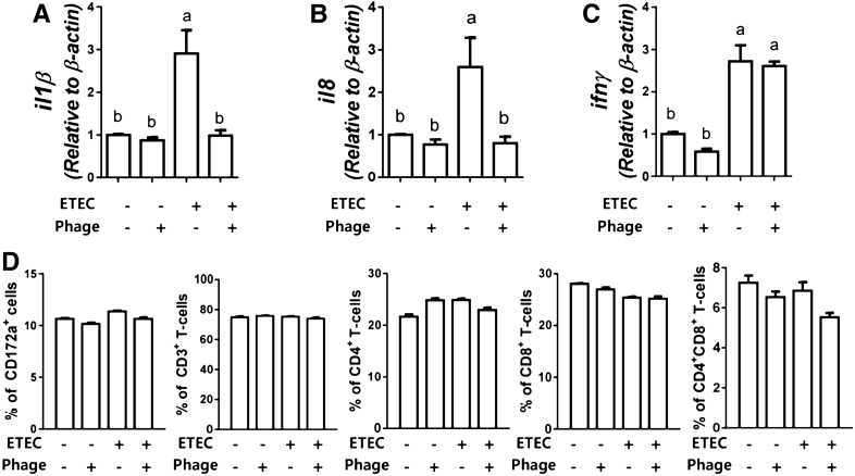
Bacteriophages, simply phages, have long been used as a potential alternative to antibiotics for livestock due to their ability to specifically kill enterotoxigenic Escherichia coli (ETEC), which is a major cause of diarrhea in piglets. However, the control of ETEC infection by phages within intestinal epithelial cells, and their relationship with host immune responses, remain poorly understood. In this study, we evaluated the effect of phage EK99P-1 against ETEC K99-infected porcine intestinal epithelial cell line (IPEC-J2). Phage EK99P-1 prevented ETEC K99-induced barrier disruption by attenuating the increased permeability mediated by the loss of tight junction proteins such as zonula occludens-1 (ZO-1), occludin, and claudin-3. ETEC K99-induced inflammatory responses, such as interleukin (IL)-8 secretion, were decreased by treatment with phage EK99P-1. We used a IPEC-J2/peripheral blood mononuclear cell (PBMC) transwell co-culture system to investigate whether the modulation of barrier disruption and chemokine secretion by phage EK99P-1 in ETEC K99-infected IPEC-J2 would influence immune cells at the site of basolateral. The results showed that phage EK99P-1 reduced the mRNA expression of ETEC K99-induced pro-inflammatory cytokines, IL-1β and IL-8, from PBMC collected on the basolateral side. Together, these results suggest that phage EK99P-1 prevented ETEC K99-induced barrier dysfunction in IPEC-J2 and alleviated inflammation caused by ETEC K99 infection. Reinforcement of the intestinal barrier, such as regulation of permeability and cytokines, by phage EK99P-1 also modulates the immune cell inflammatory response.


Conflict of interest statement

The authors declare no competing interests.

Figures

Figure 1
Figure 1
Phage EK99P-1 restored intestinal permeability in porcine intestinal epithelial cell line IPEC-J2 infected with enterotoxigenic Escherichia coli (ETEC K99). Differentiated IPEC-J2 were treated with ETEC K99 (1 × 107 cfu/mL) and phage EK99P-1 (1 × 106 pfu/mL) for 24 h. (A) Transepithelial electrical resistance (TEER) was measured in epithelial volt/ohm after 24 h of infection and (B) at the indicated time points. Each datum represents a percentage of initial TEER (n = 3). *P <0.05; **P < 0.01; ***P < 0.001. (C,D) Permeability was assessed by measuring 4- or 40-kD fluorescein isothiocyanate (FITC)-dextran transport after 24 h of infection (n = 3). Data are expressed as means ± standard deviation (SD). Means were compared by one-way analysis of variance (ANOVA), followed by the Friedman test and Tukey’s multiple comparison test. Different letters in each group indicate significant differences at P < 0.05.
Figure 2
Figure 2
Phage EK99P-1 restored the decreased expression of tight junction proteins by ETEC in IPEC-J2 infected with ETEC K99. A monolayer of confluent IPEC-J2 was treated with ETEC K99 (2 × 106 cfu/mL) and phage EK99P-1 (2 × 105 pfu/mL) for 3 h. (A) Whole-cell lysates were analyzed for the expression of ZO-1, occludin, claudin-3, and β-actin using anti-claudin-3, -occludin, -ZO-1 and -β-actin antibodies by western blot assay (n = 3). Full-length blots/gels are presented in Supplementary Fig. 4. (B) ZO-1 expression in IPEC-J2 was visualized using confocal microscopy after staining with anti-ZO-1 antibody conjugated with Alexa Fluor 488-FITC (green) and nuclei with DAPI (blue) (n = 3). Scale bar = 30 μm.
Figure 3
Figure 3
Phage EK99P-1 inhibited the adhesion of ETEC K99 to IPEC-J2. Differentiated IPEC-J2 was treated with ETEC K99 (1 × 107 cfu/mL) and phage EK99P-1 (1 × 106 pfu/mL) for 24 h. An E. coli adhesion assay was performed on IPEC-J2 (n = 3). Data are means ± SD. Means were compared using a two-tailed Student’s t-test. ****P < 0.0001 compared to the ETEC control.
Figure 4
Figure 4
Phage EK99P-1 inhibits inflammatory responses in IPEC-J2 infected with ETEC K99. Differentiated IPEC-J2 were treated with ETEC K99 (1 × 107 cfu/mL) and phage EK99P-1 (1 × 106 pfu/mL) for 24 h. (A) The expression of cytokine and chemokine mRNA in differentiated IPEC-J2 was measured by RT-PCR after 24 h of treatment (n = 3). (B) IL-8 secretion was measured at supernatant in apical and basolateral side of transwell plate by an enzyme-linked immunosorbent assay (ELISA; n = 3). Data are means ± SD. Means were compared using one-way ANOVA, followed by Friedman’s test and Tukey’s multiple comparison test. Different letters in each group indicate significant differences at P < 0.05.
Figure 5
Figure 5
Phage EK99P-1 alleviated the inflammatory response of immune cells under IPEC-J2 infection with ETEC K99. Differentiated IPEC-J2 and pPBMCs were co-cultured using transwell plates. IPEC-J2 were treated with ETEC K99 (1 × 107 cfu/mL) and phage EK99P-1 (1 × 106 pfu/mL) on the apical side for 24 h. (A–C) Expression of cytokine mRNA in pPBMC was measured by real-time quantitative RT-PCR (qRT-PCR; n = 3). (D) pPBMCs were stained with anti-CD172a, -CD3e, -CD4, or -CD8a antibodies to analyze the population of immune cells using flow cytometry. Data are mean percentages of each cell population relative to the total cells ± SD (n = 3). Means were compared using one-way ANOVA, followed by a Friedman test corrected by Tukey’s multiple comparison test. Different letters in each group indicate significant differences at P < 0.05.

References

    1. Wilson R, Francis DH. Fimbriae and enterotoxins associated with Escherichia coli serogroups isolated from pigs with colibacillosis. Am. J. Vet. Res. 1986;47(2):213–217. - PubMed
    1. Kaper JB, Nataro JP, Mobley HL. Pathogenic Escherichia coli. Nat. Rev. Microbiol. 2004;2(2):123–140. doi: 10.1038/nrmicro818. - DOI - PubMed
    1. Nagy B, Fekete PZ. Enterotoxigenic Escherichia coli (ETEC) in farm animals. Vet. Res. 1999;30(2–3):259–284. - PubMed
    1. Chen X, et al. Prevalence of serogroups and virulence factors of Escherichia coli strains isolated from pigs with postweaning diarrhoea in eastern China. Vet. Microbiol. 2004;103(1–2):13–20. doi: 10.1016/j.vetmic.2004.06.014. - DOI - PubMed
    1. Shams Z, et al. Detection of enterotoxigenic K99 (F5) and F41 from fecal sample of calves by molecular and serological methods. Comp. Clin. Pathol. 2012;21(4):475–478. doi: 10.1007/s00580-010-1122-2. - DOI - PMC - PubMed

Publication types

MeSH terms