Review
. 2022 Mar;21(3):181-200.
doi: 10.1038/s41573-021-00371-6. Epub 2022 Jan 18.

PROTAC targeted protein degraders: the past is prologue

Affiliations
Review

PROTAC targeted protein degraders: the past is prologue

Miklós Békés et al. Nat Rev Drug Discov. 2022 Mar.

Abstract

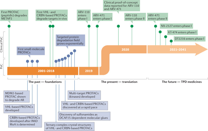
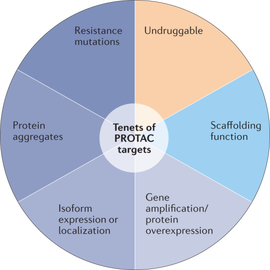
Targeted protein degradation (TPD) is an emerging therapeutic modality with the potential to tackle disease-causing proteins that have historically been highly challenging to target with conventional small molecules. In the 20 years since the concept of a proteolysis-targeting chimera (PROTAC) molecule harnessing the ubiquitin-proteasome system to degrade a target protein was reported, TPD has moved from academia to industry, where numerous companies have disclosed programmes in preclinical and early clinical development. With clinical proof-of-concept for PROTAC molecules against two well-established cancer targets provided in 2020, the field is poised to pursue targets that were previously considered 'undruggable'. In this Review, we summarize the first two decades of PROTAC discovery and assess the current landscape, with a focus on industry activity. We then discuss key areas for the future of TPD, including establishing the target classes for which TPD is most suitable, expanding the use of ubiquitin ligases to enable precision medicine and extending the modality beyond oncology.


Conflict of interest statement

C.M.C. is a consultant and shareholder in Arvinas, Inc. and Halda Therapeutics, which support research in his lab. M.B. and D.R.L. are employees and shareholders of Arvinas, Inc, which is developing drug candidates in the targeted protein degradation space.

Figures

Fig. 1
Fig. 1. The mechanism of PROTAC-mediated targeted protein degradation.
Schematic representation of the mechanism of action of proteolysis-targeted chimera (PROTAC) molecules. The PROTAC molecule (enlarged in the circle) is a heterobifunctional molecule bridging a ubiquitin ligase (in blue) and a target protein (in green). As a first step, PROTACs induce the proximity of the ligase and the substrate, such that ubiquitin (in pink) will be conjugated to the recruited substrate by the activity of the ligase. This is a catalytic step that a single PROTAC molecule can perform iteratively, enabling multiple turnover of ubiquitylation reactions, resulting in formation of ubiquitin chains on a substrate. Ubiquitin chains are then recognized by the proteasome (in red), shuttling the ubiquitylated substrate through its proteolytic chamber and degrading the target protein into small peptides (in green). Figure reproduced with permission from Arvinas, Inc.
Fig. 2
Fig. 2. Modalities in targeted protein degradation.
a | Structure and properties of two proteolysis-targeting chimeras (PROTACs) that have entered clinical trials, ARV-110 and ARV-471. PROTACs are composed of a target-binding moiety (green), a linker (orange) and an E3 ligase-binding moiety (blue). ARV-110 and ARV-471 target the androgen receptor and the oestrogen receptor, respectively, while the E3-binding ligand interacts with the cereblon (CRBN) E3 ligase. b | Schematic representation of the two foundational modalities for targeted protein degradation. Left-hand side: discovery of PROTACs composed of a target-binding moiety (green), a linker (orange) and a ligase-binding moiety (blue), enabling the rational discovery of heterobifunctional molecules to degrade a desired target. Right-hand side: opportunistic discovery of degrader molecules, whereby a known molecule is shown to have a degrader effect, making it possible to identify the E3 ligase mediating that degradation and determine whether that degrader mechanism could be expanded to target additional proteins of interest. The pros and cons of these modalities are discussed in the text and in Box 1. CMR, calculated molecular refractivity; tPSA, total polar surface area.
Fig. 3
Fig. 3. Timeline of PROTAC discoveries.
The first era of targeted protein degradation (TPD) began with publication of the pivotal proteolysis-targeting chimera (PROTAC) paper by Sakamoto et al. in 2001, which was the first demonstration of the concept that protein targets could be intentionally dragged to a ubiquitin ligase to induce their degradation using chemical tools. Between then and today, the field has grown exponentially and has moved from peptide-based tool degraders to multiple classes of fully synthetic small molecules that can induce proximity between a ligase and a protein of interest, leading to its degradation. This foundational era of TPD was capped by the first rational heterobifunctional PROTAC degrader entering clinical trials in 2019, ARV-110, which targets the androgen receptor (AR) by recruiting it to the Cullin–RING ligase 4–cereblon (CRL4–CRBN) ligase complex. The current era of TPD can be considered its initial translational phase, in which multiple molecules designed to degrade disease-causing proteins are entering the clinic with the hope of providing meaningful benefits to patients. DCAF15, DDB1- and CUL4-associated factor 15; IMiD, immunomodulatory imide drug; MoA, mechanism of action; METAP2, methionyl aminopeptidase 2; PoC, proof of concept; VHL, von Hippel–Lindau.
Fig. 4
Fig. 4. The tenets of PROTAC targets.
Proteolysis-targeting chimeras (PROTACs) bring the protein degrader function to the target; they do not need to bind within a biologically functional active site. This expands the accessible targets well beyond those that are druggable by traditional stoichiometric inhibition and provides novel ways to achieve selectivity. Proteins that may be best suited to therapeutic intervention by targeted protein degradation instead of stoichiometric inhibition include proteins with disease-causing gain of function owing to mutation, overexpression, aggregation or the differential expression or localization of protein isoforms. From a structural perspective, PROTAC targets need a small-molecule binding surface that is approachable by an E3 ligase, and ideally have an unstructured region that can be threaded into the proteasome.
Fig. 5
Fig. 5. Example Cullin–RING ligases and their substrate adaptors.
Examples of Cullin–RING ligases (CRLs) and a U-box E3 ligase modelled as complete E3–E2–ubiquitin (Ub) complexes: CRL4–cereblon (CRBN) (panel a); CRL2–von Hippel–Lindau (VHL) (panel b); CRL1–β-transducin repeat-containing E3 ubiquitin–protein ligase (β-TRCP) (panel c); CHIP (STIP1 homology and U-box-containing protein 1) (panel d). The assemblies demonstrate the different size, shape and electrostatic potential surface of each ligase, all potentially enabling degradation of a wide variety of proteins of interest when co-opted as ligases for degradation (Cullin/RING-box protein 1 (RBX1) subunits are coloured orange/purple, respectively; adaptor proteins are green; substrate receptors are blue; ligands are sea green). The surface-rendered substrate receptors in the lower images show the electrostatic potential coloured surface of the binding protein/domain of the substrate as viewed from the E2 (indicated by (>). The U-box CHIP E3 ligase is a single protein and is coloured by domain in panel d (U-box: orange; TRP, tetratricopeptide repeat: blue). The zone of ubiquitylation is defined by the reach of E2–Ub and is shown for CRL2–VHL in panel b. The high-probability ubiquitylation sites on the protein of interest are lysine residues that can be positioned within the zone and within ~4 Å of the reactive thioester between E2 and ubiquitin. The crystal structure protein databank (PDB) codes used to generate the models are: CRBN (2HYE, 3UGB, 6BN7 (ref.)), VHL (6R7F, 3UGB, 5T35 (ref.)), β-TRCP (6TTU, 3UGB) and CHIP (2C2L, 6S53 (ref.)). ELOB/ELOC, elongin B–elongin C.
Fig. 6
Fig. 6. Specialized E3 ligases for potential PROTAC applications.
a | Schematic representation of the human body, highlighting E3 ligases with increased tissue specificities that may be harnessed by targeted protein degradation (TPD) modalities to enable tissue- and cell-type-specific targeting of disease-causing proteins. b | Table showing representative E3 ligase examples from different mechanistic classes of E3 ligase, highlighting their various (but not exhaustive) attributes to be considered for proteolysis-targeting chimera (PROTAC) development, including tumour and tissue enrichment, tumour dependence (CERES/DepMap profile) and mechanistic understanding of ubiquitin ligation onto their substrates. The E3 examples were chosen for each E3 class from multiple E3-centric review articles,,,, to highlight various characteristics and the expansive choice in ligase selection for novel PROTACs. One article is referenced for each E3, but there is considerable overlap between the content of the cited references. There is no single attribute that makes or breaks an E3 ligase for PROTAC development; all attributes should be considered in totality, including the target that is being considered for degradation. CNS, central nervous system; CRBN, cereblon; DCAF2, DDB1- and CUL4-associated factor 2; ELOB, elongin B; GID4, glucose-induced degradation protein 4 homologue; KLHL40, kelch-like family member 40; MAGEs, melanoma antigen genes; RNF182, RING finger protein 182; TRIM9, tripartite motif-containing protein 9; VHL, von Hippel–Lindau.

References

    1. Surka C, et al. CC-90009, a novel cereblon E3 ligase modulator, targets acute myeloid leukemia blasts and leukemia stem cells. Blood. 2021;137:661–677. - PMC - PubMed
    1. Hansen JD, et al. Discovery of CRBN E3 ligase modulator CC-92480 for the treatment of relapsed and refractory multiple myeloma. J. Med. Chem. 2020;63:6648–6676. - PubMed
    1. Lu G, et al. The myeloma drug lenalidomide promotes the cereblon-dependent destruction of Ikaros proteins. Science. 2014;343:305–309. - PMC - PubMed
    1. Kronke J, et al. Lenalidomide causes selective degradation of IKZF1 and IKZF3 in multiple myeloma cells. Science. 2014;343:301–305. - PMC - PubMed
    1. Kronke J, et al. Lenalidomide induces ubiquitination and degradation of CK1alpha in del(5q) MDS. Nature. 2015;523:183–188. - PMC - PubMed