[Preprint]. 2022 Jan 12:2022.01.11.475918.
doi: 10.1101/2022.01.11.475918.

Shift of lung macrophage composition is associated with COVID-19 disease severity and recovery

Affiliations

Shift of lung macrophage composition is associated with COVID-19 disease severity and recovery

Steven T Chen et al. bioRxiv. .

Update in

  • A shift in lung macrophage composition is associated with COVID-19 severity and recovery.
    Chen ST, Park MD, Del Valle DM, Buckup M, Tabachnikova A, Thompson RC, Simons NW, Mouskas K, Lee B, Geanon D, D'Souza D, Dawson T, Marvin R, Nie K, Zhao Z, LeBerichel J, Chang C, Jamal H, Akturk G, Chaddha U, Mathews K, Acquah S, Brown SA, Reiss M, Harkin T, Feldmann M, Powell CA, Hook JL, Kim-Schulze S, Rahman AH, Brown BD; Mount Sinai COVID-19 Biobank Team; Beckmann ND, Gnjatic S, Kenigsberg E, Charney AW, Merad M. Chen ST, et al. Sci Transl Med. 2022 Sep 14;14(662):eabn5168. doi: 10.1126/scitranslmed.abn5168. Epub 2022 Sep 14. Sci Transl Med. 2022. PMID: 36103512 Free PMC article.

Abstract

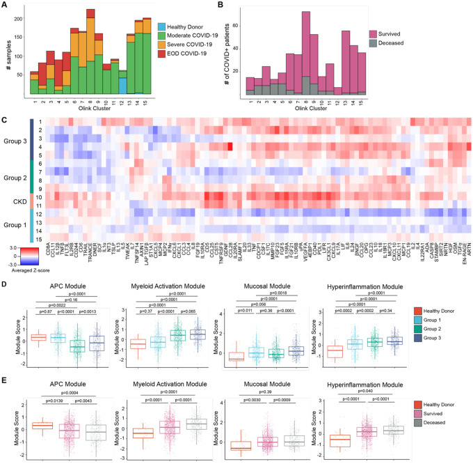
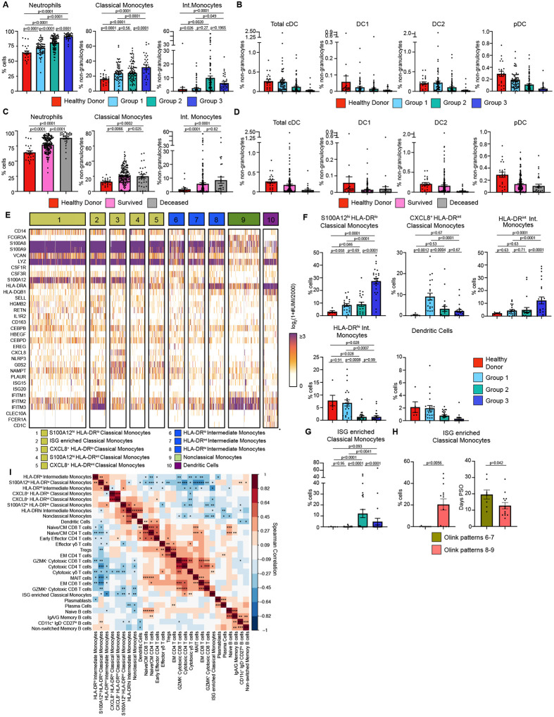
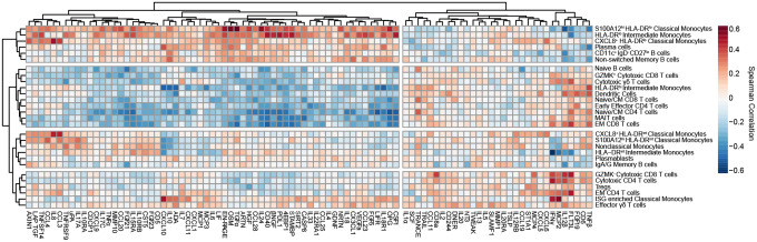
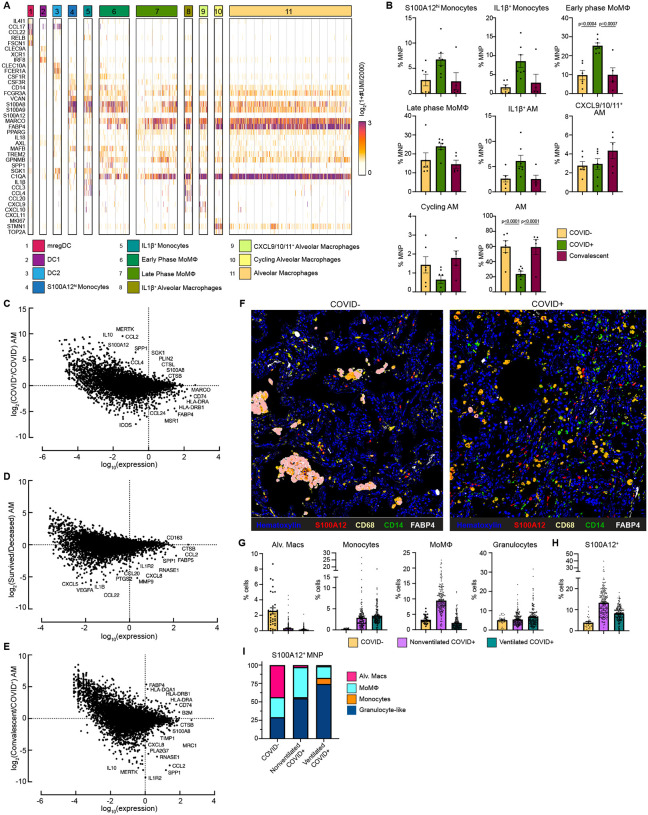
Though it has been 2 years since the start of the Coronavirus Disease 19 (COVID-19) pandemic, COVID-19 continues to be a worldwide health crisis. Despite the development of preventive vaccines, very little progress has been made to identify curative therapies to treat COVID-19 and other inflammatory diseases which remain a major unmet need in medicine. Our study sought to identify drivers of disease severity and death to develop tailored immunotherapy strategies to halt disease progression. Here we assembled the Mount Sinai COVID-19 Biobank which was comprised of ~600 hospitalized patients followed longitudinally during the peak of the pandemic. Moderate disease and survival were associated with a stronger antigen (Ag) presentation and effector T cell signature, while severe disease and death were associated with an altered Ag presentation signature, increased numbers of circulating inflammatory, immature myeloid cells, and extrafollicular activated B cells associated with autoantibody formation. Strikingly, we found that in severe COVID-19 patients, lung tissue resident alveolar macrophages (AM) were not only severely depleted, but also had an altered Ag presentation signature, and were replaced by inflammatory monocytes and monocyte-derived macrophages (MoMΦ). Notably, the size of the AM pool correlated with recovery or death, while AM loss and functionality were restored in patients that recovered. These data therefore suggest that local and systemic myeloid cell dysregulation is a driver of COVID-19 severity and that modulation of AM numbers and functionality in the lung may be a viable therapeutic strategy for the treatment of critical lung inflammatory illnesses.


Figures

Figure 1.
Figure 1.. Proteomic characterization of COVID-19 serum reveals distinct immune patterns associated with disease severity and clinical outcome.
(A) Histogram of patient samples across Olink clusters denoted by clinical severity classification. (B) Histogram of first available patient samples across Olink clusters denoted by patient projected clinical outcome. (C) Averaged z-scored heatmap of Olink inflammation panel analytes across Olink clusters. Olink clusters were grouped based on clinical severity, projected outcome, and comorbidity distribution. (D) Boxplots showing Olink module score comparisons of all Olink samples by Olink group. (E) Boxplots showing Olink module score comparisons of all Olink samples by final clinical outcome. For box plots, each dot represents a patient sample; center line, median; box limits, 25th and 75th percentile; whiskers, 1.5x interquartile range (IQR). Statistical significance (D-E) determined by 2-way ANOVA with Tukey’s Multiple Comparisons correction. Adjusted p-values shown.
Figure 2.
Figure 2.. Immature inflammatory myeloid cells associated with increased COVID-19 severity.
(A) Neutrophils (% cells), Classical Monocytes and Intermediate Monocyte frequencies (% non-granulocytes) in whole blood by Olink group measured by CyTOF. (B) DC population frequencies (% non-granulocytes) in whole blood by Olink group measured by CyTOF. (C) Neutrophils (% cells), Classical Monocytes and Intermediate Monocyte frequencies (%non-granulocytes) in whole blood by final clinical outcome measured by CyTOF. (D) DC population frequencies (% non-granulocytes) in whole blood by final clinical outcome measured by CyTOF. Conventional DC (cDC), conventional type 1 DC (DC1), conventional type 2 DC (DC2), and plasmacytoid DC (pDC) shown. (E) Heatmap showing unique molecular identifier (UMI) counts of selected genes from myeloid cell scRNAseq clusters from PBMC. (F) scRNAseq cluster cell frequencies as % of cells by Olink group (G) % of cells frequencies of ISG enriched Classical Monocytes cluster by Olink group. (H) % of cells frequencies and days PSO for ISG enriched Classical Monocytes by clusters 6–7 vs clusters 8–9. (I) Matrix of spearman correlation coefficients between identified scRNAseq PBMC clusters. *p<0.05, **p<0.005, ***p<0.0005. For bar graphs, each dot (A-D, F-H) represents a patient sample. Statistical significance (A-D, F-G) determined by 2-way ANOVA with Holm-Sidak multiple comparisons correction. Adjusted p-values shown. Statistical significance (H) determined by Mann-Whitney test.
Figure 3:
Figure 3:. Integrated analysis of scRNAseq cluster frequencies and Olink analyte abundance in serum reveals distinct immune responses to COVID-19.
Matrix of spearman correlation coefficients between identified scRNAseq PBMC clusters and Olink analyte normalized concentrations in serum. Axes ordered by hierarchical clustering.
Figure 4:
Figure 4:. Alveolar Macrophage loss and phenotypic changes in the COVID-19 lung microenvironment.
(A) Heatmap showing UMI counts of selected genes from myeloid cell scRNAseq clusters from BAL. (B) scRNAseq cluster cell frequencies as % mononuclear phagocytes (MNP) in COVID, COVID+, or convalescent patient BAL. Differential gene expression between (C) AM from COVID+ and COVID patients, (D) AM from patients that survived vs deceased, (E) AM from convalescent and COVID+ patients. (F) Overlaid, pseudocolored MICSSS image of COVID+ and COVID lungs, staining for S100A12, CD68, CD14, FABP4, and Hematoxylin. (G) Quantification of myeloid cells in MICSSS images, shown as % of cells. AM defined as FABP4+CD68+ cells; Monocytes defined as CD14+ cells; MoMΦ defined as CD14+CD68+ cells; Granulocyte-like cells defined as CD66b+ cells or by hematoxylin staining and morphology. (H) Quantification of S100A12+ cells in MICSSS images, shown as % cells in COVID patient (n=1), nonventilated COVID+ patients (n=2), or ventilated COVID+ patients (n=2). (I) Distribution of S100A12+ cells by cell type in COVID, nonventilated, or ventilated COVID+ lungs. For bar graphs, each dot represents patient sample (B) or quantification of single MICSSS region of interest (ROI) (G-H). Statistical significance (B) determined by 2-way ANOVA with Holm-Sidak multiple comparisons correction. Adjusted p-values shown.

References

    1. Charney A. W., Simons N. W., Mouskas K., Lepow L., Cheng E., Le Berichel J., Chang C., Marvin R., Del Valle D. M., Calorossi S., Lansky A., Walker L., Patel M., Xie H., Yi N., Yu A., Kang G., Liharska L. E., Moya E., Hartnett M., Hatem S., Wilkins L., Eaton M., Jamal H., Tuballes K., Chen S. T., Chung J., Harris J., Batchelor C., Lacunza J., Yishak M., Argueta K., Karekar N., Lee B., Kelly G., Geanon D., Handler D., Leech J., Stefanos H., Dawson T., Scott I., Francoeur N., Johnson J. S., Vaid A., Glicksberg B. S., Nadkarni G. N., Schadt E. E., Gelb B. D., Rahman A., Sebra R., Martin G., Agashe C., Agrawal P., Alesso-Carra K., Alibo E., Alvarez K., Amabile A., Ascolillo S., Bailey R., Begani P., Correra P. B., Brown S. A., Buckup M., Burka L., Cambron L., Carrara G., Chang S., Chien J., Chowdhury M., Bozkus C. C., Comella P., Cosgrove D., Cossarini F., Cotter L., Dave A., Dayal B., Dhainaut M., Dornfeld R., Dul K., Eber N., Elaiho C., Fabris F., Faith J., Falci D., Feng S., Fennessy B., Fernandes M., Gangadharan S., Grabowska J., Grimes G., Hamdani M., Herbinet M., Herrera E., Hochman A., Hoffman G. E., Hook J., Horta L., Humblin E., Karim S., Kim J., Lebovitch D., Lee G., Lee G. H., Lee J., Leventhal M., Lindblad K., Livanos A., Machado R., Mahmood Z., Mar K., Maskey S., Matthews P., Meckel K., Mehandru S., Mercedes C., Meyer D., Mollaoglu G., Morris S., Nie K., Nisenholtz M., Ofori-Amanfo G., Onel K., Ounadjela M., Patel V., Pruitt C., Rathi S., Redes J., Reyes-Torres I., Rodrigues A., Rodriguez A., Roudko V., Ruiz E., Scalzo P., Silva P., Schanoski A. S., Straw M., Tabachnikova S., Teague C., Upadhyaya B., Van Der Heide V., Vaninov N., Wacker D., Walsh H., Wilk C. M., Wilson J., Wilson K. M., Xue L., akomaah Yeboah N., Young S., Zaks N., Zha R., Marron T., Beckmann N., Kim-Schulze S., Gnjatic S., Merad M., Sampling the host response to SARS-CoV-2 in hospitals under siege, Nat. Med. 2020 268 26, 1157–1158 (2020). - PubMed
    1. Mantovani A., Sica A., Sozzani S., Allavena P., Vecchi A., Locati M., The chemokine system in diverse forms of macrophage activation and polarization,, doi:10.1016/j.it.2004.09.015. - DOI - PubMed
    1. Merad M., Sathe P., Helft J., Miller J., Mortha A., The Dendritic Cell Lineage: Ontogeny and Function of Dendritic Cells and Their Subsets in the Steady State and the Inflamed Setting, Annu. Rev. Immunol. 31, 563–604 (2013). - PMC - PubMed
    1. Locksley R. M., Killeen N., Lenardo M. J., The TNF and TNF Receptor Superfamilies: Integrating Mammalian Biology, Cell 104, 487–501 (2001). - PubMed
    1. DeLuca L. S., Ng D., Gao Y., Wortzman M. E., Watts T. H., Gommerman J. L., LTβR signaling in dendritic cells induces a type I IFN response that is required for optimal clonal expansion of CD8+ T cells, Proc. Natl. Acad. Sci. U. S. A. 108, 2046–2051 (2011). - PMC - PubMed

Publication types