. 2022 Jan;27(7):074710.
doi: 10.1117/1.JBO.27.7.074710.

Kernel Flow: a high channel count scalable time-domain functional near-infrared spectroscopy system

Affiliations

Kernel Flow: a high channel count scalable time-domain functional near-infrared spectroscopy system

Han Y Ban et al. J Biomed Opt. 2022 Jan.

Abstract

Significance: Time-domain functional near-infrared spectroscopy (TD-fNIRS) has been considered as the gold standard of noninvasive optical brain imaging devices. However, due to the high cost, complexity, and large form factor, it has not been as widely adopted as continuous wave NIRS systems.

Aim: Kernel Flow is a TD-fNIRS system that has been designed to break through these limitations by maintaining the performance of a research grade TD-fNIRS system while integrating all of the components into a small modular device.

Approach: The Kernel Flow modules are built around miniaturized laser drivers, custom integrated circuits, and specialized detectors. The modules can be assembled into a system with dense channel coverage over the entire head.

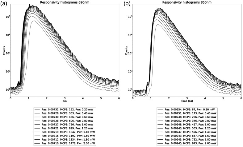
Results: We show performance similar to benchtop systems with our miniaturized device as characterized by standardized tissue and optical phantom protocols for TD-fNIRS and human neuroscience results.

Conclusions: The miniaturized design of the Kernel Flow system allows for broader applications of TD-fNIRS.

Keywords: functional near-infrared spectroscopy; optical brain imaging; optical properties; single-photon detectors; time-resolved spectroscopy; tissue optics.


Figures

Fig. 1
Fig. 1
Side view of the modules and structural plates that make up the Flow system. Each plate of modules establishes a fixed distance between the sources and detectors of the modules within the plate. The spacing between plates can be controlled using spacers that should be adjusted based on the user’s head size and desired regions of interest.
Fig. 2
Fig. 2
An exploded view showing the details of all of the module subassemblies.
Fig. 3
Fig. 3
Overall architecture of the Flow hardware. A global clock is distributed from the primary MCU subassembly to all module endpoints.
Fig. 4
Fig. 4
Laser subassembly showing the two different wavelengths of edge-emitting lasers, which are pulsed into a silver-coated prism to combine them into the same source light pipe. The PCB assembly is secured to an aluminum base that holds the prism in place and also serves as a heat sink for the laser diodes.
Fig. 5
Fig. 5
Photos of the detector subassembly showing the six Kernel custom detectors (front) and associated support circuitry (back).
Fig. 6
Fig. 6
Module cross section showing the optical configuration of one of the Flow modules. The center light pipe captures the light from the two lasers (690 and 850 nm) after it is reflected off of the prism. The source light leaves the module through a 3.1-mm aperture (red arrow). The detector optics are indicated by the blue arrows and are used to image the tip of the detector light pipes onto the custom designed detectors.
Fig. 7
Fig. 7
Schematic of setup used for the BIP responsivity measurement.
Fig. 8
Fig. 8
(a) Schematic of the custom fixture used to collect an IRF with the Kernel Flow module. (b) Photo of the IRF fixture.
Fig. 9
Fig. 9
Top view schematic of the depth contrast measurement. A 5-mm diameter × 5-mm height black PVC cylinder target is measured using a single source and detector channel between two Kernel Flow modules with a 31.2-mm separation. The target is moved at different depths, at which the contrast and CNR are calculated with respect to the homogeneous liquid phantom (with μa=0.01  mm1 and μs=1.0  mm1 at 690 nm).
Fig. 10
Fig. 10
Subset of the MEDPHOT phantoms with their absorption (rows) and scattering (columns) properties. For each row/column, the average value of the absorption/scattering is shown at the two wavelengths at which the Kernel Flow system operates: 690 (red values) and 850 nm (black values) (units are cm1).
Fig. 11
Fig. 11
Distribution of times of flight (DTOF) of laser light transmitted through the BIP phantom for the responsivity measurement at 690 and 850 nm. Histograms with increasing counts correspond with increasing power, count rate, and responsivity.
Fig. 12
Fig. 12
Histogram of the IRF measurement for 690 and 850 nm from a 10-mm separation source-detector pair.
Fig. 13
Fig. 13
Plots of the 70-min-long IRF stability measurements showing (a) the total counts of the histogram over time for 690- (red) and 850-nm (black) measurements for a representative module (shown are mean ± standard deviation across all detectors); (b) the FWHM showing the width variation of the histograms for the module shown in (a) over time, (c) the change in the mean ToF for the module shown in (a). (d) Total counts (top), FWHM (middle), and mean ToF (bottom) for all within-module detectors for 690-nm laser (23 modules are shown). (e) Same as in (d) but for 850-nm laser.
Fig. 14
Fig. 14
The contrast and CNR of the photon counts at 690 nm for time windows of 500 ps (starting at the rising edge of the histogram), as a 100-mm3 volume black PVC cylinder is sequentially placed at depths from 6 to 30 mm. The histograms are from a single source-detector pair between two Kernel modules with a distance of 31.2 mm.
Fig. 15
Fig. 15
(a) and (b) The nominal μa and μs value of the 12 phantoms (blue x’s) and the estimated values derived from measurements by the Flow module, at 690 nm. Each 5-ms histogram generates an independent measurement, plotted in black with an opacity of α=0.01. Thus, pale gray means few histograms result in estimates at those values, whereas black indicates dozens to hundreds of estimates at that value. Thin black lines connect the nominal value to the median of estimates, to guide the eye. (a) 690-nm results and (b) 850 nm. (c) Nominal μa versus estimated μa. Correlation coefficients are 0.988 and 0.993 for 690 and 850 nm, respectively. (d) Nominal μs versus estimated μs. Correlation coefficients are 0.989 and 0.984 for 690 and 850 nm.
Fig. 16
Fig. 16
Estimated μa values of the absorption coefficient across time for the two different wavelengths using recordings from a Flow module (solid lines; red: 690 nm, gray: 850 nm). Dashed horizontal lines indicate true values and shaded areas correspond to the ±5% of the true values. Note that the recovered μa values were smoothed using a 1-min Gaussian smoothing kernel.
Fig. 17
Fig. 17
Plot showing total counts for six detectors on the forehead of a participant.
Fig. 18
Fig. 18
(a) The mean total counts per histogram versus the source-detector distance. (b) The number of channels that pass quality control checks at each SDS.
Fig. 19
Fig. 19
Hemodynamic responses in a finger-tapping task. (a) Time course of hemodynamic responses (HbO: red and HbR: blue) during an entire finger-tapping session from participant one. Data are from a representative channel in the right motor cortex. Note the increase in HbO signal during the left- (green) and decrease in HbO signal during the right- (purple) tapping blocks. (b) Shown are the epoched hemodynamic responses (mean ± standard error across blocks) from participant two for the example channels marked in the topographic plot (middle; light blue and orange circles correspond to the location of the channels shown in the left and right panels, respectively). Topographic plot demonstrates the results of cluster-based permutation tests that were used to assess a significant difference between the responses (HbO) during left- and right-tapping conditions. Colorbar values correspond to log10(p) multiplied by the direction of the effect. The p-values were corrected to account for multiple comparisons. (c) Same as (b) but for data from another finger-tapping session with participant two.

References

    1. Torricelli A., et al. , “Time domain functional NIRS imaging for human brain mapping,” NeuroImage 85, 28–50 (2014).NEIMEF10.1016/j.neuroimage.2013.05.106 - DOI - PubMed
    1. Wojtkiewicz S., et al. , “Self-calibrating time-resolved near infrared spectroscopy,” Biomed. Opt. Express 10(5), 2657 (2019).BOEICL10.1364/BOE.10.002657 - DOI - PMC - PubMed
    1. Wabnitz H., et al. , “Depth-selective data analysis for time-domain fNIRS: moments vs time windows,” Biomed. Opt. Express 11(8), 4224 (2020).BOEICL10.1364/BOE.396585 - DOI - PMC - PubMed
    1. Zhang C., et al. , “A CMOS SPAD imager with collision detection and 128 dynamically reallocating TDCs for single-photon counting and 3D time-of-flight imaging,” Sensors 18(11), 4016 (2018).SNSRES10.3390/s18114016 - DOI - PMC - PubMed
    1. Jiang J., et al. , “2.5 Hz sample rate time-domain near-infrared optical tomography based on SPAD-camera image tissue hemodynamics,” Biomed. Opt. Express 13(1), 133 (2022).BOEICL10.1364/BOE.441061 - DOI - PMC - PubMed

MeSH terms