. 2022 Mar 23;96(6):e0164121.
doi: 10.1128/JVI.01641-21. Epub 2022 Jan 19.

Contribution of RNA-RNA Interactions Mediated by the Genome Packaging Signals for the Selective Genome Packaging of Influenza A Virus

Affiliations

Contribution of RNA-RNA Interactions Mediated by the Genome Packaging Signals for the Selective Genome Packaging of Influenza A Virus

Sho Miyamoto et al. J Virol. .

Abstract

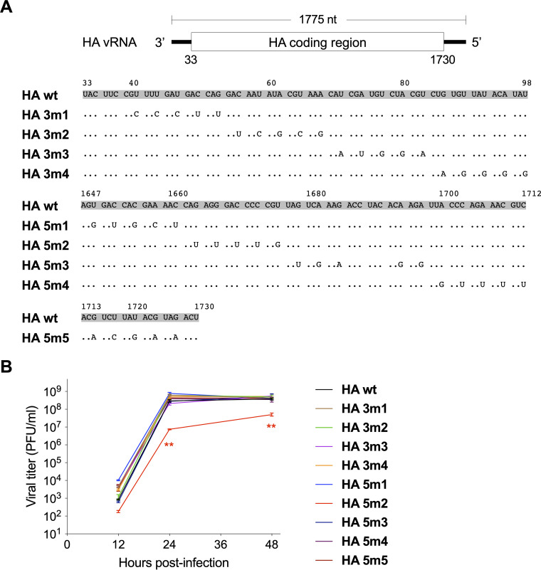
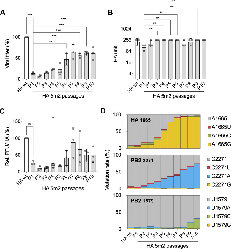
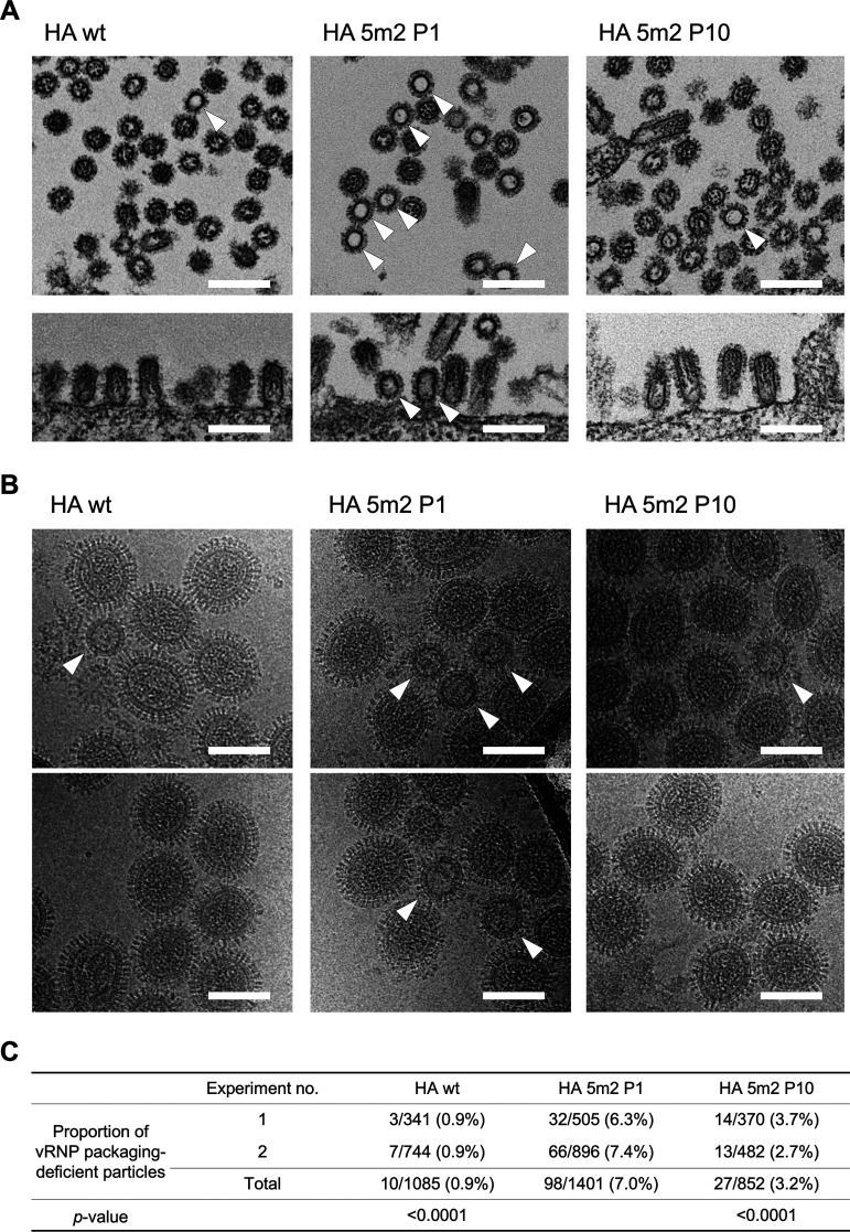
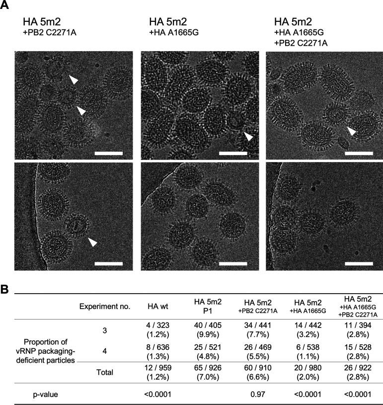
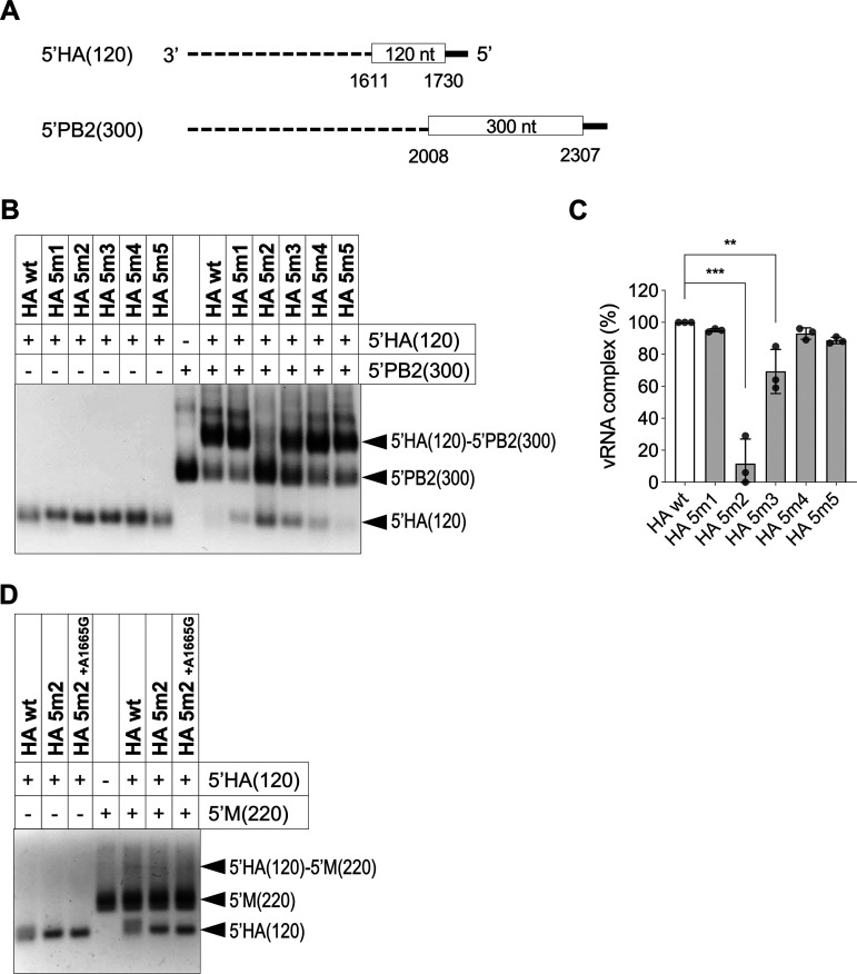
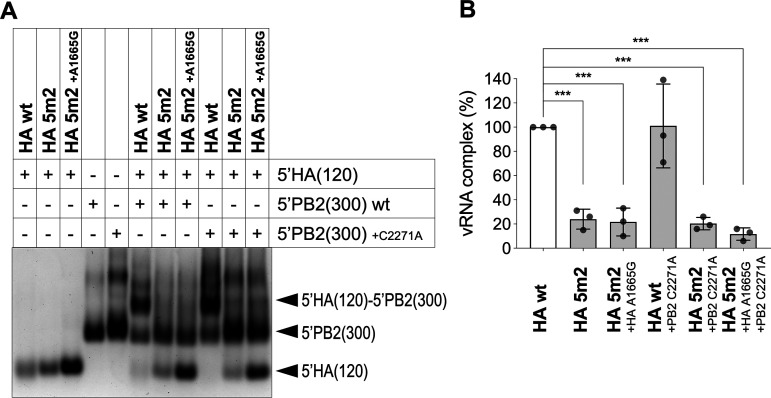
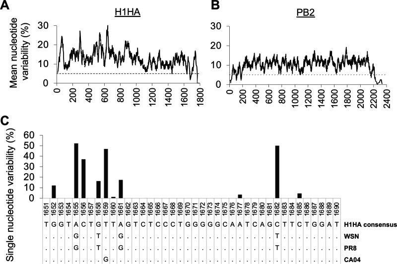
The influenza A virus genome is composed of eight single-stranded negative-sense viral RNA segments (vRNAs). The eight vRNAs are selectively packaged into each progeny virion. This process likely involves specific interactions between the vRNAs via segment-specific packaging signals located in both the 3'- and 5'-terminal regions of the respective vRNAs. To assess the importance of vRNA-vRNA interactions via packaging signals for selective genome packaging, we generated mutant viruses possessing silent mutations in the packaging signal region of the hemagglutinin (HA) vRNA. A mutant virus possessing silent mutations in nucleotides (nt) 1664 to 1676 resulted in defects in HA vRNA incorporation and showed a reduction in viral growth. After serial passage, the mutant virus acquired additional mutations in the 5'-terminal packaging signal regions of both the HA and polymerase basic 2 (PB2) vRNAs. These mutations contributed to the recovery of viral growth and HA vRNA packaging efficiency. In addition, an RNA-RNA interaction between the 5' ends of HA and PB2 vRNAs was confirmed in vitro, and this interaction was disrupted following the introduction of silent mutations in the HA vRNA. Thus, our results demonstrated that RNA-RNA interactions between the packaging signal regions of HA vRNA and PB2 vRNA are important for selective genome packaging. IMPORTANCE While numerous viral genomes comprise a single genome segment, the influenza A virus possesses eight segmented genomes. Influenza A virus can benefit from having a segmented genome because the segments can reassort with other strains of the influenza virus to create new genetically distinct strains. The influenza A virus efficiently incorporates one copy of each of its eight genomic segments per viral particle. However, the mechanism by which each segment is specifically selected is poorly understood. The genome segments contain RNA signals that facilitate the incorporation of segments into virus particles. These regions may facilitate specific interactions between the genome segments, creating an eight-segment complex, which can then be packaged into individual particles. In this study, we provide evidence that RNA signals contribute to specific interactions between two of the influenza virus genome segments.

Keywords: genome; genome packaging; influenza virus.


Conflict of interest statement

The authors declare no conflict of interest.

Figures

FIG 1
FIG 1
Generation of mutant influenza A viruses by reverse genetics and replication efficiencies of these mutant viruses. (A) Schematic diagram of mutant HA vRNAs with silent mutations introduced into the packaging signal sequences. The 3′ and 5′ ends of the HA coding region (nt 33 to 98 and nt 1649 to 1730, respectively) are shown in the vRNA sequence. (B) Growth of mutant HA viruses in MDCK cells. 293T cells were transfected with plasmids to produce the mutant and HA wt viruses. MDCK cells were infected with these mutant and HA wt viruses at an MOI of 10−5. Virus yields at 12 hpi, 24 hpi, and 48 hpi were determined by a plaque assay in MDCK cells. The data represent the geometric mean ± SD of three independent experiments. Viral titers at each time point were compared with that of the HA wt virus by using one-way ANOVA with Dunnett’s test; **, P < 0.01.
FIG 2
FIG 2
Analysis of passaged HA 5m2 virus growth and infectivity. (A) Viral growth of passaged HA 5m2 viruses. The HA 5m2 virus was passaged in MDCK cells at an MOI of 10−5. For each passage, supernatants were collected at 48 hpi, and viral yields were determined by plaque assay in MDCK cells. (B and C) HA units (B) and PFU/HA ratio (C) of the passaged HA 5m2 viruses. The viral titer and the PFU/HA ratio were compared with those of the wt HA virus. Assays were performed in triplicate for technical replicates. The viral titer and PFU/HA ratio are represented as the mean ± SD. HA units represent the geometric mean ±SD. (D) Mutations introduced during serial passaging of the HA 5m2 virus. The mutation rates of the HA wt and HA 5m2 P1 to P10 viruses are shown. Peak values for each nucleotide were measured using Sanger sequencing and the SnapGene software.
FIG 3
FIG 3
Analysis of mutant HA 5m2 virus growth and infectivity. (A) Viral growth of mutant HA 5m2 viruses in low-MOI infections. 293T cells were transfected with plasmids to produce the mutant HA5m2 and HA wt viruses. After infection of MDCK cells at an MOI of 10−5, viral yields were determined at 12 hpi, 24 hpi, and 48 hpi. Data represent the geometric mean ± SD of three independent experiments. Viral titers at each time point were compared with that of HA 5m2 P1 virus using one-way ANOVA with Dunnett’s test; ***, P < 0.001 (versus HA 5m2 +PB2 C2271A [green] and HA 5m2 +HA A1665G +PB2 C2271A [orange] at 12 hpi; versus HA wt [black], HA 5m2 +HA A1665G +PB2 C2271A, and HA 5m2 P10 [blue] at 24 and 48 hpi). (B and C) HA units (B) and PFU/HA ratio (C) of the mutant HA 5m2 viruses in low-MOI infections. (D) Viral growth of mutant HA 5m2 viruses in high-MOI single-cycle infections. MDCK cells were infected at an MOI of 1. The supernatants were harvested at 10 hpi. (E and F) HA units (E) and PFU/HA ratio (F) of the mutant HA 5m2 viruses in high-MOI single-cycle infections. Viral titers and HA units represent the geometric mean ± SD of the three independent biological replicates. The PFU/HA ratio represents the mean ± SD of the three independent experiments. Data were compared with that of HA 5m2 P1 virus using one-way ANOVA with Tukey’s test; *, P < 0.05; **, P < 0.01; ***, P < 0.001.
FIG 4
FIG 4
Effect of the additional mutations in HA 5m2 P10 virus on HA and PB2 protein function. (A) HA protein amounts incorporated into progeny virions. MDCK cells were infected with the viruses at a multiplicity of infection (MOI) of 1. At 10 hpi, the supernatant was harvested and purified. The purified viruses were analyzed by Western blotting using an anti-influenza A virus polyclonal antibody against HA, NP, and M1. (B) Ratio of NP to M1 protein in the purified viruses. The NP/M1 ratio represents the mean ± SD of three independent experiments. Data were compared using one-way ANOVA. (C) Effect of silent and additional mutations on HA expression. RNPs possessing HA vRNA were reconstructed in 293 cells. At 36 h posttransfection (hpt), cell lysate was analyzed by Western blotting. Representative images from two independent experiments are shown. (D) Replication and transcription efficiencies of viral polymerases. MDCK cells were infected with the viruses at an MOI of 1. Total RNA was extracted at 6 hpi. HA vRNA, HA cRNA, and HA mRNA were analyzed using strand-specific RT-qPCR analysis. The data represent the geometric mean ± SD of three independent experiments for biological replicates. Copy numbers of each RNA strand among the indicated viruses were compared using one-way ANOVA.
FIG 5
FIG 5
Packaging of individual vRNA segments into progeny viral particles. (A) Relative amounts of eight vRNAs from the HA wt, HA 5m2 P10, HA 5m2 P1, and mutant HA 5m2 viruses in low-MOI infections. MDCK cells were infected with the viruses at an MOI of 10−5. The supernatants were collected at 48 hpi and purified. The amount of vRNA extracted from purified virus particles was quantified by RT-qPCR. (B) Relative amounts of eight vRNA from the viruses in high-MOI single-cycle infections. MDCK cells were infected at an MOI of 1. The supernatants were harvested at 10 hpi. All vRNAs were normalized to the amount of PB1 vRNA and to the average of that contained in the HA wt virus. Data represent the mean ± SD from two independent experiments, each conducted in triplicate. The amounts of individual vRNAs were compared with that in HA 5m2 P1 virus using one-way ANOVA with Dunnett’s test; *, P < 0.05; **, P < 0.01; ***, P < 0.001.
FIG 6
FIG 6
Morphology of HA 5m2 P1, HA5m2 P10, and HA wt viruses. (A) Ultrathin sections of viral particles budding from infected MDCK cells were observed using transverse sectioning (upper panel) and longitudinal sectioning (lower panel). Empty particles are indicated by arrowheads. Bars, 200 nm. (B) The purified virus particles were observed by cryo-EM. vRNP packaging-deficient particles are indicated by arrowheads. Bars, 100 nm. (C) Effect of silent mutations and passages on the packaging of vRNPs into progeny virions. Proportions of vRNP packaging-deficient particles in each virus were compared with that in the HA 5m2 P1 virus using one-way ANOVA with Dunnett’s test.
FIG 7
FIG 7
Morphology of the mutant HA 5m2 viruses. (A) Purified virus particles were observed by cryo-EM. vRNP packaging-deficient particles are indicated by arrowheads. Bars, 100 nm. (B) Effect of the additional mutations on the packaging of vRNPs into progeny virions. Proportions of vRNP packaging-deficient particles in each virus were compared with that of the HA 5m2 P1 virus using one-way ANOVA with Dunnett’s test.
FIG 8
FIG 8
RNA-RNA interaction between 5′-end sequences of HA and PB2 vRNAs. (A) Schematic representation of 5'HA(120) and 5'PB2(300) vRNAs. 5'HA(120) and 5'PB2(300) contain 120 or 300 nucleotides of the coding regions (white box) with the noncoding regions (solid line) at their 5′ ends, respectively. The dashed line represents the deleted regions. (B) The effect of silent mutations in 5'HA(120) vRNA on binding to 5'PB2(300) vRNA was analyzed by a gel shift assay. 5'PB2(300) vRNA was incubated with HA wt and mutated 5'HA(120) vRNAs as indicated at the top of the gel image. Individual vRNA bands and vRNA complexes are indicated on the right. Respective band intensities of input 5'HA(120) mutants were 85% to 140% of that of HA wt. (C) Quantification of 5'HA(120)-5'PB2(300) complexes for each lane. The band intensity of the complex is indicated relative to that of the wild type. The data represent the mean ± SD of three independent experiments. Dunnett’s test; **, P < 0.01; ***, P < 0.001. (D) Specificity of the RNA-RNA interaction with the 5′ end of HA vRNA with mutations. Interactions of 5′HA(120) and 5'M(220) vRNAs were analyzed by a gel shift assay. Combinations of 5'HA(120) and 5'M(220) vRNAs are indicated at the top of the gel image.
FIG 9
FIG 9
RNA-RNA interaction of 5′-end sequences of HA and PB2 vRNAs with mutations. (A) The effect of the additional mutations in 5'HA(120) and 5'PB2(300) vRNAs was analyzed by a gel shift assay. Combinations of 5'HA(120) and 5'PB2(300) vRNAs are indicated at the top of the gel image. Respective band intensities of input 5'HA(120) mutants were 100% to 132% of those of HA wt. (B) Quantification of 5'HA(120)-5'PB2(300) complexes for each lane. The band intensity of the complex is indicated relative to that of the wild type. The data represent the mean ± SD of three independent experiments. Dunnett’s test; ***, P < 0.001.
FIG 10
FIG 10
Nucleotide variabilities of H1HA and PB2 genome segments. Nucleotide variabilities of 100 H1N1 sequences and several HA subtype sequences are shown for the HA and PB2 segment, respectively. (A-B) A sliding window algorithm of 25 nucleotides was used to generate variability plots. The coding regions showing a mean nucleotide variability of less than 5% were as follows; HA nt 1445, 1633 to 1636, and 1669 to 1696; PB2 nt 25 to 39, 101 to 113, 121 to 122, 172 to 185, 2156 to 2159, 2165 to 2166, and 2219 to 2307. (C) Single nucleotide variability of the H1HA nt 1651 to 1690 region. PR8, A/Puerto Rico/8/1934 (H1N1); CA04, A/California/04/2009 (H1N1).
FIG 11
FIG 11
Predicted RNA structure of HA nt 1649 to 1682 and PB2 nt 2256 to 2308 regions. (A) RNA secondary structures in the HA nt 1649 to 1682 vRNA in HA wt, HA 5m2, HA 5m2 +A1665G variant. The nucleotides of point mutations in HA 5m2 are represented by red circles. An additional mutation, A1665G (vRNA strand sequence from the 3′ end), is represented by an orange circle. (B) Structure in the PB2 nt 2256 to 2308 vRNA in PB2 wt and PB2 +C2271A. An additional mutation, C2271A, is represented by a red circle. (C) Schematic diagram of HA vRNA interaction region with PB2 vRNA. Upper, sequences of the interaction region previously reported by Dadonaite et al. (26) (green). Lower, map of predicted RNA structure and RNA-RNA interaction sites in the 5′ end of HA vRNA and PB2 vRNA. Green lines represent interaction regions with HA and PB2 vRNAs, respectively. The additional mutations introduced during passage of HA 5m2 virus are indicated.

Similar articles

Cited by

References

    1. Williams GD, Townsend D, Wylie KM, Kim PJ, Amarasinghe GK, Kutluay SB, Boon ACM. 2018. Nucleotide resolution mapping of influenza A virus nucleoprotein-RNA interactions reveals RNA features required for replication. Nat Commun 9:465. 10.1038/s41467-018-02886-w. - DOI - PMC - PubMed
    1. Lee N, Le Sage V, Nanni AV, Snyder DJ, Cooper VS, Lakdawala SS. 2017. Genome-wide analysis of influenza viral RNA and nucleoprotein association. Nucleic Acids Res 45:8968–8977. 10.1093/nar/gkx584. - DOI - PMC - PubMed
    1. Fujii K, Fujii Y, Noda T, Muramoto Y, Watanabe T, Takada A, Goto H, Horimoto T, Kawaoka Y. 2005. Importance of both the coding and the segment-specific noncoding regions of the influenza A virus NS segment for its efficient incorporation into virions. J Virol 79:3766–3774. 10.1128/JVI.79.6.3766-3774.2005. - DOI - PMC - PubMed
    1. Fujii Y, Goto H, Watanabe T, Yoshida T, Kawaoka Y. 2003. Selective incorporation of influenza virus RNA segments into virions. Proc Natl Acad Sci USA 100:2002–2007. 10.1073/pnas.0437772100. - DOI - PMC - PubMed
    1. Muramoto Y, Takada A, Fujii K, Noda T, Iwatsuki-Horimoto K, Watanabe S, Horimoto T, Kida H, Kawaoka Y. 2006. Hierarchy among viral RNA (vRNA) segments in their role in vRNA incorporation into influenza A virions. J Virol 80:2318–2325. 10.1128/JVI.80.5.2318-2325.2006. - DOI - PMC - PubMed

Publication types