Review
. 2022 May;36(13-15):844-863.
doi: 10.1089/ars.2021.0145. Epub 2022 Apr 18.

Mitochondrial Fusion, Fission, and Mitophagy in Cardiac Diseases: Challenges and Therapeutic Opportunities

Affiliations
Review

Mitochondrial Fusion, Fission, and Mitophagy in Cardiac Diseases: Challenges and Therapeutic Opportunities

Débora da Luz Scheffer et al. Antioxid Redox Signal. 2022 May.

Abstract

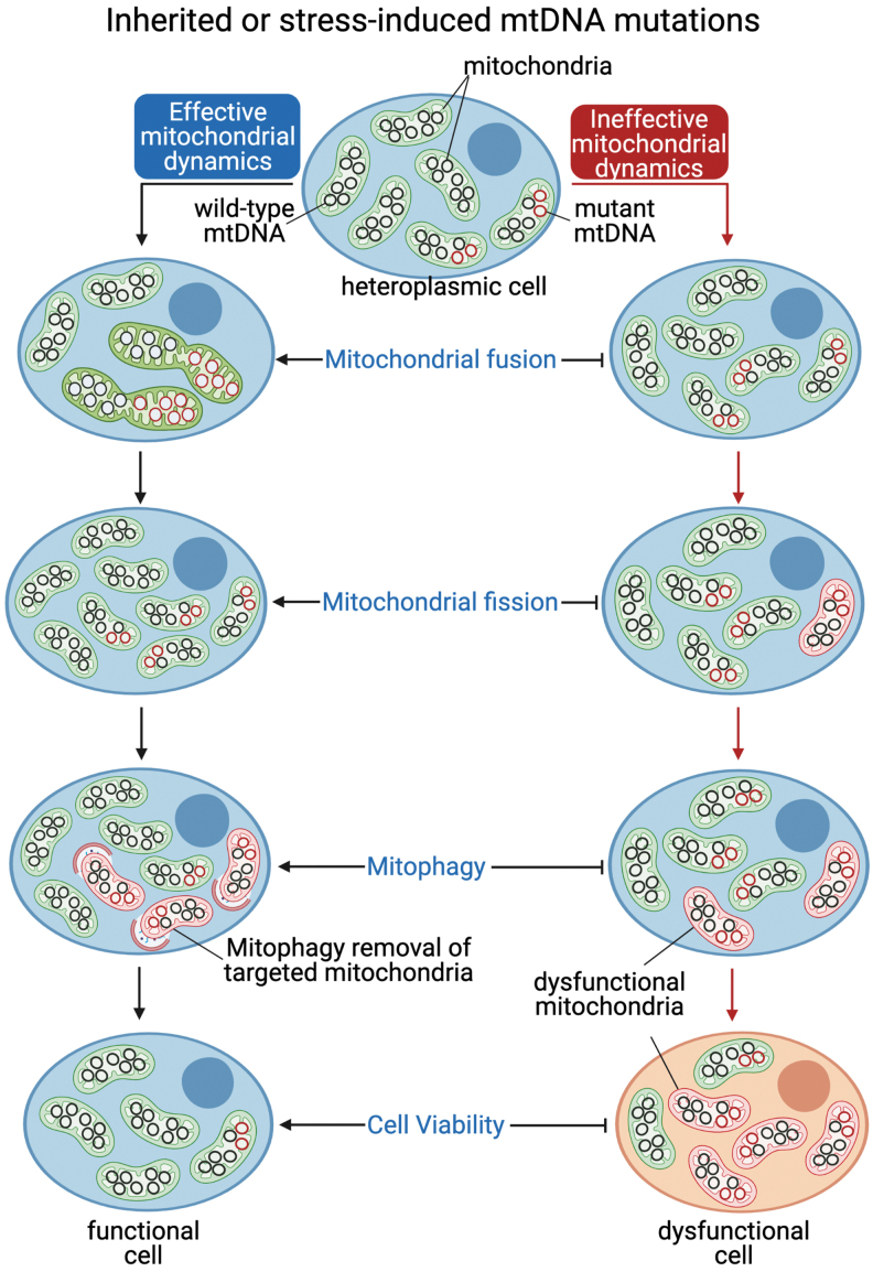
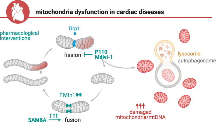
Significance: Mitochondria play a critical role in the physiology of the heart by controlling cardiac metabolism, function, and remodeling. Accumulation of fragmented and damaged mitochondria is a hallmark of cardiac diseases. Recent Advances: Disruption of quality control systems that maintain mitochondrial number, size, and shape through fission/fusion balance and mitophagy results in dysfunctional mitochondria, defective mitochondrial segregation, impaired cardiac bioenergetics, and excessive oxidative stress. Critical Issues: Pharmacological tools that improve the cardiac pool of healthy mitochondria through inhibition of excessive mitochondrial fission, boosting mitochondrial fusion, or increasing the clearance of damaged mitochondria have emerged as promising approaches to improve the prognosis of heart diseases. Future Directions: There is a reasonable amount of preclinical evidence supporting the effectiveness of molecules targeting mitochondrial fission and fusion to treat cardiac diseases. The current and future challenges are turning these lead molecules into treatments. Clinical studies focusing on acute (i.e., myocardial infarction) and chronic (i.e., heart failure) cardiac diseases are needed to validate the effectiveness of such strategies in improving mitochondrial morphology, metabolism, and cardiac function. Antioxid. Redox Signal. 36, 844-863.

Keywords: bioenergetic dysfunction; cardiovascular diseases; metabolism; mitochondrial dynamics; oxidative stress; therapy.


Conflict of interest statement

Patents on the design and application of mitochondrial fission peptide inhibitors have been filed. The authors claim that there is no conflict of interest related to this work.

Figures

FIG. 1.
FIG. 1.
Proposed model for impaired mitochondrial bioenergetics and redox balance in cardiac diseases based on previous studies (32, 73, 164), which focused on the interplay between NAD(P)H/NAD(P)+ redox state and mitochondrial ROS generation. 4-HNE, 4-hydroxynonenal; α-KG, α-ketoglutarate; GPX, glutathione peroxidase; GR, glutathione reductase; GSH/GSSG, reduced/oxidized glutathione; H2O2, hydrogen peroxide; IDPm, mitochondrial NADP+-dependent isocitrate dehydrogenase; MCU, mitochondrial Ca2+ uniporter; MDA, malondialdehyde; MEP, malic enzyme; mNCE, Na+/Ca2+ exchanger; Mn-SOD, Mn2+-dependent superoxide dismutase; O2, anion superoxide; OH, hydroxyl radical; ONOO, peroxynitrite; PRX, peroxiredoxin; ROS, reactive oxygen species; TCA, tricarboxylic acid; TR, thioredoxin reductase; TRXr/o, reduced/oxidized thioredoxin. Created with BioRender.com
FIG. 2.
FIG. 2.
Proposed model for mitochondrial fission and fusion. The mitochondrial fission is promoted by Drp1, a cytosolic GTPase that translocates to the mitochondria upon activation and binds to specific proteins located at the outer mitochondrial membrane, including mitochondrial Fis1, Mff, MiD49, and MiD51 (37). Drp1-Mff and Drp1-Fis1 interaction induces two distinct mitochondrial fission signatures and plays a critical role in the mitochondrial physiology and pathology (106). In addition, Mff, MiD49, and MiD51 are essential for Drp1-dependent mitochondrial scission and release of MDV (109). Mitochondrial fusion is coordinated by mitofusins (Mfn1 and Mfn2), located at the outer mitochondrial membrane, and the Opa1, located at the inner mitochondrial membrane. Mitofusins contain a GTPase domain involved in the hydrolysis of GTP, which is required for the mitofusin oligomerization and fusion of the outer mitochondrial membranes of adjacent mitochondria. Drp1, dynamin-related protein 1; Fis1, fission 1 protein; GTPase, guanosine triphosphatase; MDV, mitochondrial-derived vesicles; Mff, mitochondrial fission factor; Mfn1, mitofusin 1; Mfn2, mitofusin 2; MiD49, mitochondrial dynamics protein of 49 kDa; MiD51, mitochondrial dynamics protein of 51 kDa; Opa1, optic atrophy factor 1.
FIG. 3.
FIG. 3.
Mitochondrial fission/fusion imbalance and impaired mitophagy result in defective mitochondrial dynamics, which negatively affects critical processes in cardiac physiology such as ATP production, redox balance, mtDNA segregation, mitochondrial-ER tethering, and mitochondrial and cytosolic calcium handling. mtDNA, mitochondrial DNA; ER, endoplasmic reticulum.
FIG. 4.
FIG. 4.
Proposed model for mitochondrial mitophagy. (A) Mitophagy selectively targets dysfunctional mitochondria for removal and is closely linked to mitochondrial fission and fusion (147). PINK1-Parkin-mediated mitophagy is triggered by mitochondrial depolarization, which results in PINK1 accumulation in the outer mitochondrial membrane and the subsequent phosphorylation/recruitment of Parkin (a cytosolic E3-ubiquitin ligase) to the mitochondrial surface (133, 171). (B) Parkin-mediated ubiquitination of outer mitochondrial membrane proteins results in the recruitment of autophagy adaptors (p62, OPTN, and NDP52) and initiates autophagosome formation through LC3 binding and the subsequent lysosomal degradation of autophagosome engulfed mitochondria (127, 140). PINK1, PTEN-induced kinase 1.
FIG. 5.
FIG. 5.
Proposed model for mitochondrial segregation under accumulation of inherited or stress-mediated mtDNA mutations. Coexistence of wild-type (black circle) and mutated (red circle) mtDNA in the same cell is defined as mtDNA heteroplasmy, directly regulated and selected by constant events of mitochondrial fission, fusion, and mitophagy (142, 192). Defective mitochondrial dynamics results in accumulation of mtDNA mutations, mitochondrial dysfunction, and the consequent reduction of cell viability (117).
FIG. 6.
FIG. 6.
Proposed model for therapeutics targeting mitochondrial dynamics. Impaired mitochondrial fission/fusion and mitophagy and the consequent accumulation of fragmented and dysfunctional mitochondria are common hallmarks of cardiac diseases. Pharmacological interventions that improve mitochondrial connectiveness and clearance such as P110, Mdivi-1, or SAMβA play a critical role in the maintenance of mitochondrial health, therefore becoming potential novel therapeutic targets for cardiac diseases. SAMβA, selective antagonist of Mfn1-βIIPKC association.

References

    1. Abeliovich H, Zarei M, Rigbolt KTG, Youle RJ, and Dengjel J. Involvement of mitochondrial dynamics in the segregation of mitochondrial matrix proteins during stationary phase mitophagy. Nat Commun 4: 2789, 2013. - PMC - PubMed
    1. Abrahams JP, Leslie AGW, Lutter R, and Walker JE. Structure at 2.8 Â resolution of F1-ATPase from bovine heart mitochondria. Nature 370: 621–628, 1994. - PubMed
    1. Acín-Pérez R, Fernández-Silva P, Peleato ML, Pérez-Martos A, and Enriquez JA. Respiratory active mitochondrial supercomplexes. Mol Cell 32: P529–P539, 2008. - PubMed
    1. Acin-Perez R, Lechuga-Vieco AV, DelMarMuñoz M, Nieto-Arellano R, Torroja C, Sánchez-Cabo F, Jiménez C, González-Guerra A, Carrascoso I, Benincá C, Quiros PM, López-Otín C, Castellano JM, Ruíz-Cabello J, Jiménez-Borreguero LJ, and Enríquez JA. Ablation of the stress protease OMA1 protects against heart failure in mice. Sci Transl Med 10: eaan4935, 2018. - PubMed
    1. Amati-Bonneau P, Valentino ML, Reynier P, Gallardo ME, Bornstein B, Boissière A, Campos Y, Rivera H, De La Aleja JG, Carroccia R, Iommarini L, Labauge P, Figarella-Branger D, Marcorelles P, Furby A, Beauvais K, Letournel F, Liguori R, La Morgia C, Montagna P, Liguori M, Zanna C, Rugolo M, Cossarizza A, Wissinger B, Verny C, Schwarzenbacher R, Martín MÁ, Arenas J, Ayuso C, Garesse R, Lenaers G, Bonneau D, and Carelli V. OPA1 mutations induce mitochondrial DNA instability and optic atrophy “plus” phenotypes. Brain 131: 338–351, 2008. - PubMed

Publication types