. 2022 Feb 4;21(2):494-506.
doi: 10.1021/acs.jproteome.1c00865. Epub 2022 Jan 19.

Interrogating Kinase-Substrate Relationships with Proximity Labeling and Phosphorylation Enrichment

Affiliations

Interrogating Kinase-Substrate Relationships with Proximity Labeling and Phosphorylation Enrichment

Tian Zhang et al. J Proteome Res. .

Abstract

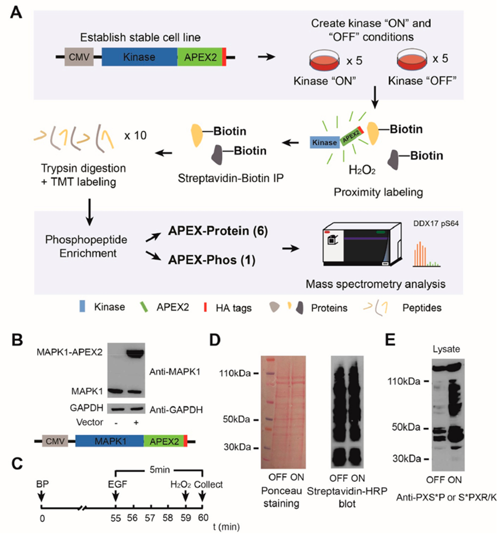
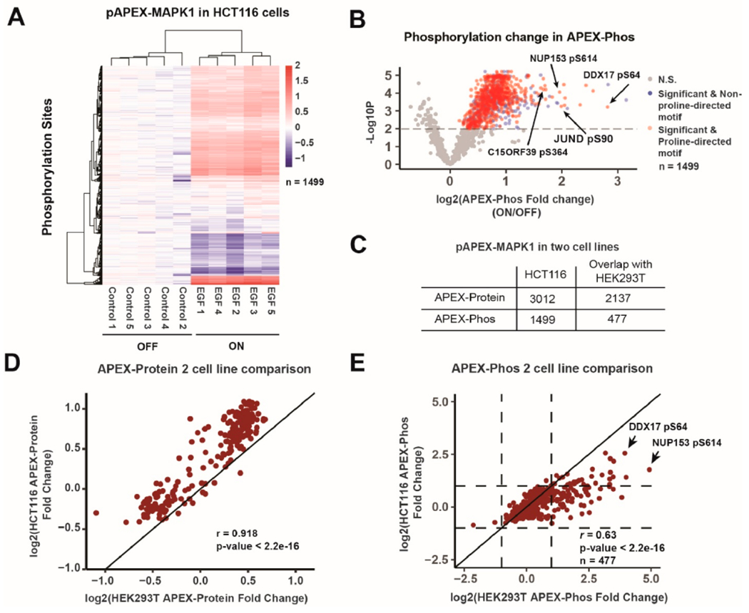
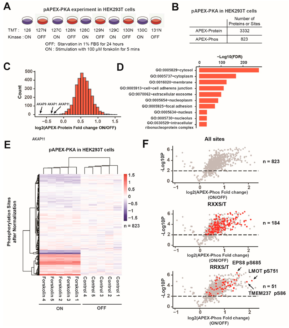
Kinases govern many cellular responses through the reversible transfer of a phosphate moiety to their substrates. However, pairing a substrate with a kinase is challenging. In proximity labeling experiments, proteins proximal to a target protein are marked by biotinylation, and mass spectrometry can be used for their identification. Here, we combine ascorbate peroxidase (APEX) proximity labeling and a phosphorylation enrichment-based workflow, Phospho-APEX (pAPEX), to rapidly identify phosphorylated and biotinylated neighbor proteins which can be considered for candidate substrates. The pAPEX strategy enriches and quantifies differences in proximity for proteins and phosphorylation sites proximal to an APEX2-tagged kinase under the kinase "ON" and kinase "OFF" conditions. As a proof of concept, we identified candidate substrates of MAPK1 in HEK293T and HCT116 cells and candidate substrates of PKA in HEK293T cells. In addition to many known substrates, C15orf39 was identified and confirmed as a novel MAPK1 substrate. In all, we adapted the proximity labeling-based platform to accommodate phosphorylation analysis for kinase substrate identification.

Keywords: MAPK1; PKA; kinases; phosphopeptide enrichment; proximity labeling; substrates identification.


Conflict of interest statement

The authors declare no competing financial interest.

Figures

Figure 1.
Figure 1.
Phospho-APEX (pAPEX) enriches proteins in close proximity to APEX2 tagged kinase. (A) Workflow of a pAPEX experiment. Cells are engineered to stably express the targeted kinase with an APEX2 tag and 3× HA epitope tags. Proximity labeling is performed under the kinase “OFF” and kinase “ON” conditions with replicates. During 1 min of H2O2 treatment, APEX2 catalyzes the transfer of biotin phenol (BP) to proteins surroundings the APEX2 fused protein radius (1–10 nm). In a typical TMT-10plex experiment, five replicates of cells under the kinase “ON” and “OFF” conditions are used to improve the statistical power. Biotinylated proteins are pulled down by streptavidin immunoprecipitation, digested, TMT-labeled, and combined prior to phosphopeptide enrichment. The flow-through of the phosphopeptide enrichment is fractionated into six fractions, which we refer to as APEX-Protein. The phosphopeptide fraction and six fractions of APEX-Protein are analyzed using mass spectrometry with reporter ion quantification. Comparing phosphopeptide abundance changes for the kinase “ON” and “OFF” conditions can reveal candidate kinase substrates. (B) Western blotting analysis showing the stable expression of the MAPK1-APEX2 fusion protein in HEK293T cells. (C) Time course of the proximity labeling experiment under the kinase “ON” condition in MAPK1-pAPEX experiment. (D) Biotinylated proteins from the proximity labeling experiment in HEK293T cells were analyzed by Ponceau staining (left) and streptavidin-HRP Western blotting (right). (E) Western blotting analysis using antibodies recognizing the consensus motif for MAPK phosphorylation.
Figure 2.
Figure 2.
pAPEX quantified proteins and phosphorylated peptides in close proximity to APEX2 tagged MAPK1 in HEK293T cells. (A) Experimental overview. (B) Proteins and sites identified. (C) Biotinylated proteins are highly enriched in nuclear proteins after 5 min EGF treatment, suggesting relocalization of MAPK1. (D) Gene ontology enrichment categories for proteins quantified in APEX-Protein analysis using proteins highlighted in red or blue in Panel C. Heatmap (E) and volcano plot (F) of phosphorylation sites quantified. In the heatmap, log2(ON/OFF) ratios were calculated and plotted; the five control and five EGF-treated samples clustered. In the volcano plot, sites are colored based on significance and whether or not they contained a proline-directed motif. (G) Pie chart of the significant (colored) sites in Panel F. The majority (84%) of sites were classified as proline-directed.
Figure 3.
Figure 3.
APEX quantified proteins and phosphorylated peptides in close proximity to APEX2 tagged MAPK1 in HCT116 cells. The experiment shown in Figure 2A was repeated in HCT116 cells. Heatmap (A) and volcano plot (B) of phosphorylation sites quantified in pAPEX-MAPK1 in HCT116 cells. (C) Comparison of proteins and phosphorylation sites quantified in the two cell lines. (D) Scatterplot of the APEX-Protein change in the two cell lines (r = 0.918). The unity line is drawn. (E) Scatterplot of APEX-Phos changes in the two cell lines (r = 0.63). HEK293T cells are more responsive on average to EGF treatment than HCT116 cells which harbor the KRASG12D allele already activating the MAPK pathway. The unity line is drawn. Dashed vertical and horizontal lines represent 0 (no change) and 1 (2-fold change). pS614 of NUP153 and pS64 of DDX17 are indicated.
Figure 4.
Figure 4.
pAPEX quantified proteins and phosphorylated peptides in close proximity to APEX2 tagged PKA in HEK293T cells. (A) Experimental overview. (B) Quantified proteins and sites. (C) Distribution of log2(fold change ON/OFF) for proteins quantified in the APEX-Protein. (D) Gene ontology enrichment categories for all proteins quantified in the APEX-Protein analysis. (E) Heatmap of phosphorylation sites quantified. Log2(fold change (ON/OFF)) were calculated and plotted. The five control and five forskolin treated samples clustered. (F) Volcano plots of quantified phosphorylation events. Sites with significant changes are colored based on site’s underlying motif (RXXS/T or RRXS/T).
Figure 5.
Figure 5.
C15orf39 can be phosphorylated by MAPK1 in vitro. (A) Phosphorylation levels for several sites in two different cell lines. T364 from C15orf39 increased in both HCT116 and HEK293T cells after EGF treatment. (B) Epitope-tagged C15orf39 localizes to the nucleus. HEK293T cells stably expressing C-terminal FLAG-HA tagged (CTAP) C15orf39 were fixed and stained with anti-HA antibodies to visualize ectopic protein localization via confocal microscopy. Max intensity projections of 2 μm z-stacks are shown; scale bar represents 20 μm. (C) HA immunoprecipitation of C15orf39 specifically enriched MAD2L1 as a candidate binding partner. HSPA5 is shown as a background control. (D) In vitro kinase assay using MAPK1 and purified C15orf39 protein. After incubation with ATP and MAPK1, C15orf39 showed increased abundance of the phosphorylated form of LEPPLpTPR and decreased signal for the nonphosphorylated form (LEPPLTPR). Base peak chromatograms using isotopic envelope peaks were visualized in Skyline.

References

    1. Manning G; Whyte DB; Martinez R; Hunter T; Sudarsanam S The Protein Kinase Complement of the Human Genome. Science 2002, 298, 1912. - PubMed
    1. Wang Z; Cole PA Catalytic mechanisms and regulation of protein kinases. Methods Enzymol 2014, 548, 1–21. - PMC - PubMed
    1. Grundke-Iqbal I; et al. Abnormal phosphorylation of the microtubule-associated protein tau (tau) in Alzheimer cytoskeletal pathology. Proc. Natl. Acad. Sci. U. S. A 1986, 83, 4913–4917. - PMC - PubMed
    1. Lahiry P; Torkamani A; Schork NJ; Hegele RA Kinase mutations in human disease: interpreting genotype-phenotype relationships. Nat. Rev. Genet 2010, 11, 60–74. - PubMed
    1. Bhullar KS; Lagaron NO; McGowan EM; Parmar I; Jha A; Hubbard BP; Rupasinghe HPV. Kinase-targeted cancer therapies: progress, challenges and future directions. Mol. Cancer 2018, 17,48–48. - PMC - PubMed

Publication types

Substances