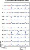
Hybrid machine learning to localize atrial flutter substrates using the surface 12-lead electrocardiogram
- PMID: 35045172
- PMCID: PMC9301972
- DOI: 10.1093/europace/euab322
Hybrid machine learning to localize atrial flutter substrates using the surface 12-lead electrocardiogram
Abstract
Aims: Atrial flutter (AFlut) is a common re-entrant atrial tachycardia driven by self-sustainable mechanisms that cause excitations to propagate along pathways different from sinus rhythm. Intra-cardiac electrophysiological mapping and catheter ablation are often performed without detailed prior knowledge of the mechanism perpetuating AFlut, likely prolonging the procedure time of these invasive interventions. We sought to discriminate the AFlut location [cavotricuspid isthmus-dependent (CTI), peri-mitral, and other left atrium (LA) AFlut classes] with a machine learning-based algorithm using only the non-invasive signals from the 12-lead electrocardiogram (ECG).
Methods and results: Hybrid 12-lead ECG dataset of 1769 signals was used (1424 in silico ECGs, and 345 clinical ECGs from 115 patients-three different ECG segments over time were extracted from each patient corresponding to single AFlut cycles). Seventy-seven features were extracted. A decision tree classifier with a hold-out classification approach was trained, validated, and tested on the dataset randomly split after selecting the most informative features. The clinical test set comprised 38 patients (114 clinical ECGs). The classifier yielded 76.3% accuracy on the clinical test set with a sensitivity of 89.7%, 75.0%, and 64.1% and a positive predictive value of 71.4%, 75.0%, and 86.2% for CTI, peri-mitral, and other LA class, respectively. Considering majority vote of the three segments taken from each patient, the CTI class was correctly classified at 92%.
Conclusion: Our results show that a machine learning classifier relying only on non-invasive signals can potentially identify the location of AFlut mechanisms. This method could aid in planning and tailoring patient-specific AFlut treatments.
Keywords: Atrial flutter; Cardiac modelling; Electrocardiography; Machine learning; Personalized medicine.
© The Author(s) 2022. Published by Oxford University Press on behalf of the European Society of Cardiology.
Figures


References
-
- Granada J, Uribe W, Chyou P-H, Maassen K, Vierkant R, Smith PNet al. . Incidence and predictors of atrial flutter in the general population. J Am Coll Cardiol 2000;36:2242–6. - PubMed
-
- Page RL, Joglar JA, Caldwell MA, Calkins H, Conti JB, Deal BJet al. ; Evidence Review Committee Chair‡ . 2015 ACC/AHA/HRS guideline for the management of adult patients with supraventricular tachycardia: a report of the American College of Cardiology/American Heart Association Task Force on Clinical Practice Guidelines and the Heart Rhythm Society. Circulation 2016;133:e506–74. - PubMed
-
- Saoudi N, Cosío F, Waldo A, Chen SA, Iesaka Y, Lesh Met al. ; Working Group of Arrhythmias of the European of Cardiology and the North American Society of Pacing and Electrophysiology . A classification of atrial flutter and regular atrial tachycardia according to electrophysiological mechanisms and anatomical bases; a Statement from a Joint Expert Group from The Working Group of Arrhythmias of the European Society of Cardiology and the North American Society of Pacing and Electrophysiology. Eur Heart J 2001;22:1162–82. - PubMed
-
- Pappone C, Oreto G, Rosanio S, Vicedomini G, Tocchi M, Gugliotta Fet al. . Atrial electroanatomic remodeling after circumferential radiofrequency pulmonary vein ablation. Circulation 2001;104:2539–44. - PubMed

