. 2022 Mar:165:1-9.
doi: 10.1016/j.lungcan.2022.01.003. Epub 2022 Jan 6.

Image-guided interventional radiological delivery of chimeric antigen receptor (CAR) T cells for pleural malignancies in a phase I/II clinical trial

Affiliations

Image-guided interventional radiological delivery of chimeric antigen receptor (CAR) T cells for pleural malignancies in a phase I/II clinical trial

Mario Ghosn et al. Lung Cancer. 2022 Mar.

Abstract

Objectives: We describe techniques and results of image-guided delivery of mesothelin-targeted chimeric antigen receptor (CAR) T cells in patients with pleural malignancies in a phase I/II trial (ClinicalTrials.gov: NCT02414269).

Materials and methods: Patients without a pleural catheter or who lack effusion for insertion of a catheter (31 of 41) were administered intrapleural CAR T cells by interventional radiologists under image guidance by computed tomography or ultrasound. CAR T cells were administered through a needle in an accessible pleural loculation (intracavitary) or following an induced loculated artificial pneumothorax. In patients where intracavitary infusion was not feasible, CAR T cells were injected via percutaneous approach either surrounding and/or in the pleural nodule/thickening (intratumoral). Pre- and post-procedural clinical, laboratory, and imaging findings were assessed.

Results: CAR T cells were administered intrapleurally in 31 patients (33 procedures, 2 patients were administered a second dose) with successful delivery of planned dose (10-186 mL); 14/33 (42%) intracavitary and 19/33 (58%) intratumoral. All procedures were completed within 2 h of T-cell thawing. There were no procedure-related adverse events greater than grade 1 (1 in 3 patients had prior ipsilateral pleural fusion procedures). The most common imaging finding was ground glass opacities with interlobular septal thickening and/or consolidation, observed in 12/33 (36%) procedures. There was no difference in the incidence of fever, CRP, IL-6, and peak vector copy number in the peripheral blood between infusion methods.

Conclusion: Image-guided intrapleural delivery of CAR T cells using intracavitary or intratumoral routes is feasible, repeatable and safe across anatomically variable pleural cancers.

Keywords: Adoptive cell therapy; Malignant pleural effusion; Malignant pleural mesothelioma; Pleural metastases; Regional delivery.


Figures

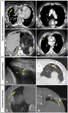
Figure 1.
Figure 1.. Various clinical presentations and image-guided intracavitary and intratumoral infusion routes of CAR T cells for pleural malignancies.
(A) Contrast enhanced chest CT showing diffuse right pleural thickening, with multiple nodules of various sizes (arrowheads). Note the extension of the pleural thickening to the interlobar fissure (long arrow). (B) Contrast enhanced chest CT showing a single right pleural mass (arrowhead). (C) This patient presented with only a small right pleural effusion (arrowhead). (D) The most challenging disease presentations were patients with only minimal pleural thickening (arrowhead), here measured at 9 mm thickness. (E) CAR T cells were injected into the pleural space (arrowhead), as shown in this case (Patient 37) where a 5Fr Yueh needle was inserted (arrow) under ultrasound guidance. (F) Axial non-contrast chest CT scan (Patient 39) after a successful pleural expansion showing the artificially induced pneumothorax (arrowhead) and a catheter placed within the newly expanded pleural cavity (arrow). (G) Axial non contrast chest CT scan (Patient 13) showing direct intratumoral infusion with a 17G needle (arrow) inserted percutaneously from an anterior intercostal approach directly into a right pleural mass (arrowhead) that was extending to the fissure. (H) Axial non contrast chest CT scan (Patient 19) showing another intratumoral infusion target, here a 12mm pleural thickening (arrowhead), also targeted with a 17G needle (arrow) using a posterior approach.
Figure 2.
Figure 2.. Summarized technical details for image guided regional delivery of CAR T cells.
Bar chart showing injected volume, type of target and material used for each procedure. Cases of pleural expansion were noted.
Figure 3.
Figure 3.. Intra- and post-procedural imaging characteristics.
(A) Attempted pleural expansion in Patient 25 who had prior pleurectomy decortication and radiation therapy with a 19G needle (short arrow) to facilitate pleural infusion of CAR T cells was not successful. Air administered through the needle tracked superiorly and along the right pectoralis muscle (long arrow). Thus, infusion was performed directly into a pleural nodule (arrowhead). (B) In this same patient, increasing ground glass opacities and interlobular septal thickening (arrowhead) were observed on intraprocedural non contrast chest axial CT scan. Note the absence of these parenchymal findings before the infusion, as shown in A. The patient respiratory status was stable throughout without cough or change in oxygen saturation suggesting intralymphatic/visceral pleural administration. Following successful administration of planned volume, the 19G needle was then removed and post procedure radiographs showed stability of the small right pneumothorax (long arrow), not requiring any chest tube placement. (C) Axial CT scan of the chest performed 3 weeks after CAR T cells infusion showed complete resolution of previous imaging findings. (D) In another example, pre-procedural CT scan of the chest (Patient 19*) performed 5 days before the procedure showed normal lung parenchyma in the right lower lobe. (E) Mild ground glass opacities and interlobular septal thickening (arrowhead) occurred after pleural injection of CAR T cells. (F) CT of the chest performed 1 month after CAR T cells infusion showed near complete resolution of previous imaging findings. (G) Slight extravasation of infusate below the diaphragm (long arrows) occurred in one patient (Patient 41), without associated clinical symptoms. (H) Table summarizing per procedural and post infusion imaging findings. Ground glass opacities and interlobular septal thickening were mild to moderate in all cases except in Patient 25 where they were diffuse. IR= interventional radiology
Figure 4.
Figure 4.. Algorithmic approach for optimal CAR T-cell infusion route selection.

References

    1. Maude SL, Laetsch TW, Buechner J, Rives S, Boyer M, Bittencourt H, Bader P, Verneris MR, Stefanski HE, Myers GD, Qayed M, De Moerloose B, Hiramatsu H, Schlis K, Davis KL, Martin PL, Nemecek ER, Yanik GA, Peters C, Baruchel A, Boissel N, Mechinaud F, Balduzzi A, Krueger J, June CH, Levine BL, Wood P, Taran T, Leung M, Mueller KT, Zhang Y, Sen K, Lebwohl D, Pulsipher MA, Grupp SA, Tisagenlecleucel in children and young adults with B-cell lymphoblastic leukemia, N Engl J Med 378(5) (2018) 439–448. 10.1056/NEJMoa1709866 - DOI - PMC - PubMed
    1. Park JH, Riviere I, Gonen M, Wang X, Senechal B, Curran KJ, Sauter C, Wang Y, Santomasso B, Mead E, Roshal M, Maslak P, Davila M, Brentjens RJ, Sadelain M, Long-term follow-up of CD19 CAR therapy in acute lymphoblastic leukemia, N Engl J Med 378(5) (2018) 449–459. 10.1056/NEJMoa1709919 - DOI - PMC - PubMed
    1. Schuster SJ, Svoboda J, Chong EA, Nasta SD, Mato AR, Anak Ö, Brogdon JL, Pruteanu-Malinici I, Bhoj V, Landsburg D, Wasik M, Levine BL, Lacey SF, Melenhorst JJ, Porter DL, June CH, Chimeric antigen receptor T cells in refractory B-cell lymphomas, N Engl J Med 377(26) (2017) 2545–2554. 10.1056/NEJMoa1708566 - DOI - PMC - PubMed
    1. Adusumilli PS, Cherkassky L, Villena-Vargas J, Colovos C, Servais E, Plotkin J, Jones DR, Sadelain M, Regional delivery of mesothelin-targeted CAR T cell therapy generates potent and long-lasting CD4-dependent tumor immunity, Sci Transl Med 6(261) (2014) 261ra151. 10.1126/scitranslmed.3010162 - DOI - PMC - PubMed
    1. Maus MV, Haas AR, Beatty GL, Albelda SM, Levine BL, Liu X, Zhao Y, Kalos M, June CH, T cells expressing chimeric antigen receptors can cause anaphylaxis in humans, Cancer Immunol Res 1 (2013) 26–31. 10.1158/2326-6066 - DOI - PMC - PubMed

Associated data