

Ultrasonographic atlas of splenic lesions
- PMID: 35045593
- PMCID: PMC8942726
- DOI: 10.14366/usg.21189
Ultrasonographic atlas of splenic lesions
Abstract
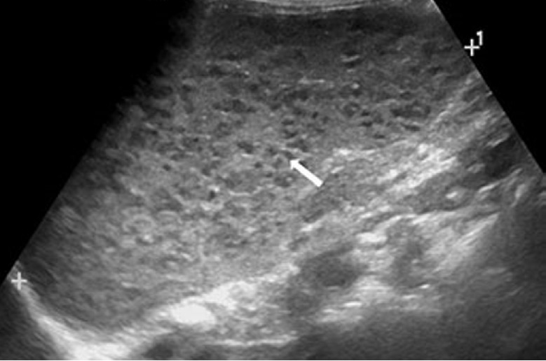
Ultrasonography (US) is widely used for abdominal imaging. Its noninvasiveness, extensive range of application, and low cost make US a useful and valuable tool for the detection, diagnosis, and follow-up of splenic abnormalities. Concomitantly with the increasing frequency of imaging, more splenic lesions are being discovered and the requirements for the differential diagnosis are rising. In this pictorial essay, we introduce the representative US findings of many different splenic lesions, including normal sonographic findings, normal variants and congenital anomalies, infectious conditions, benign and malignant neoplasms, and non-neoplastic lesions. Knowledge of the US features of various splenic lesions will help narrow the differential diagnosis and guide clinical decision-making.
Keywords: Spleen; Splenic diseases; Splenic infarction; Splenic neoplasms; Ultrasonography.
Conflict of interest statement
No potential conflict of interest relevant to this article was reported.
Figures
References
-
- Freeman SJ. In: Clinical ultrasound. 3rd ed. Allan PL, Baxter GM, Weston MJ, editors. Edinburgh: Churchill Livingstone; 2011. Spleen; pp. 324–347.
-
- Thipphavong S, Duigenan S, Schindera ST, Gee MS, Philips S. Nonneoplastic, benign, and malignant splenic diseases: cross-sectional imaging findings and rare disease entities. AJR Am J Roentgenol. 2014;203:315–322. - PubMed
-
- Benter T, Kluhs L, Teichgraber U. Sonography of the spleen. J Ultrasound Med. 2011;30:1281–1293. - PubMed
LinkOut - more resources
Full Text Sources
