. 2022 Jan 25;119(4):e2019161119.
doi: 10.1073/pnas.2019161119.

ISM1 protects lung homeostasis via cell-surface GRP78-mediated alveolar macrophage apoptosis

Affiliations

ISM1 protects lung homeostasis via cell-surface GRP78-mediated alveolar macrophage apoptosis

Terence Y W Lam et al. Proc Natl Acad Sci U S A. .

Abstract

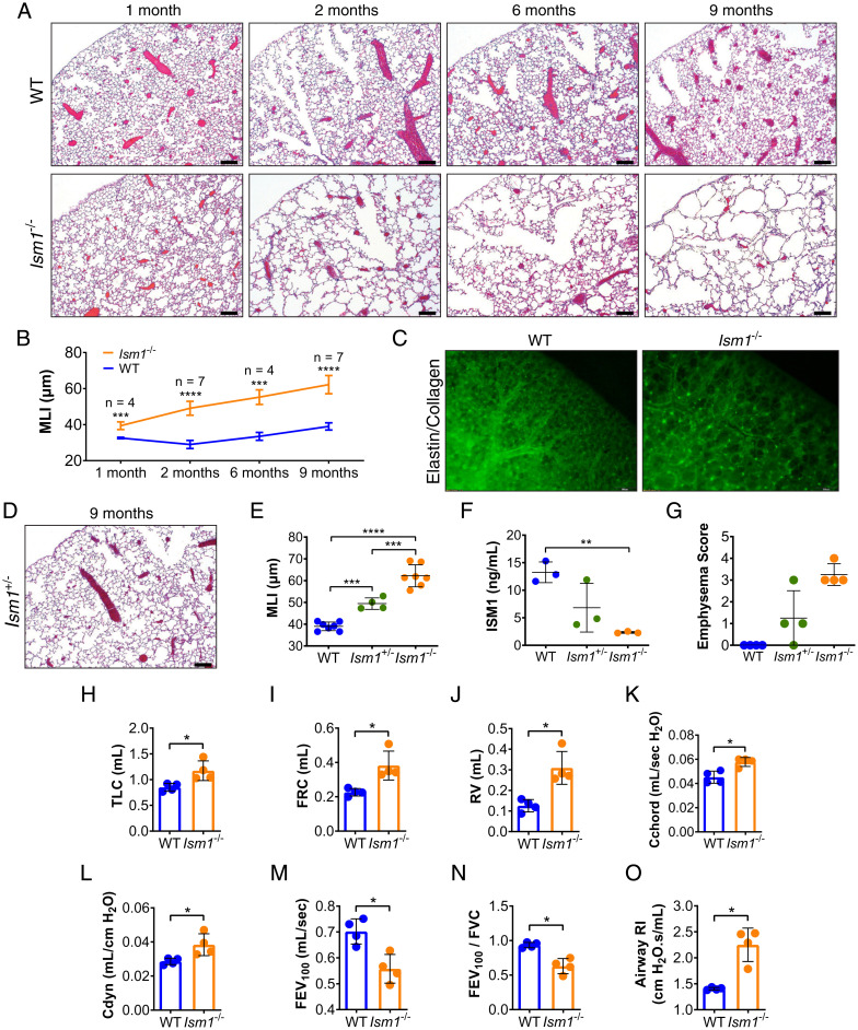
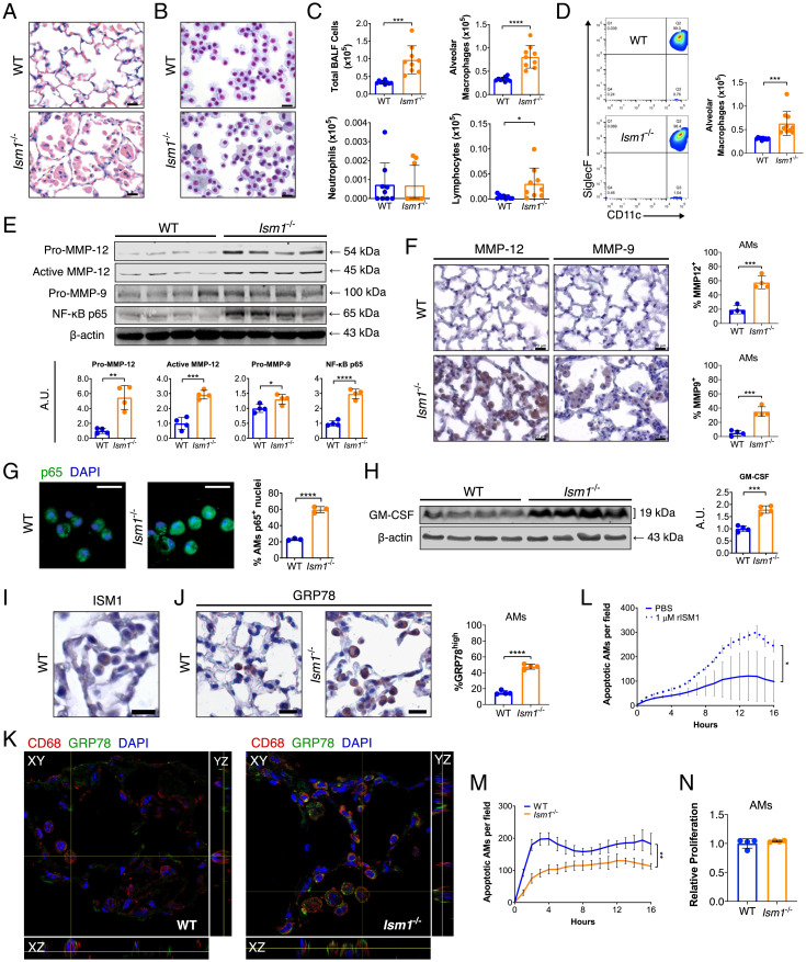
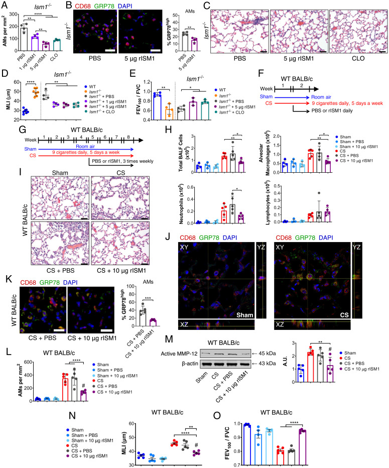
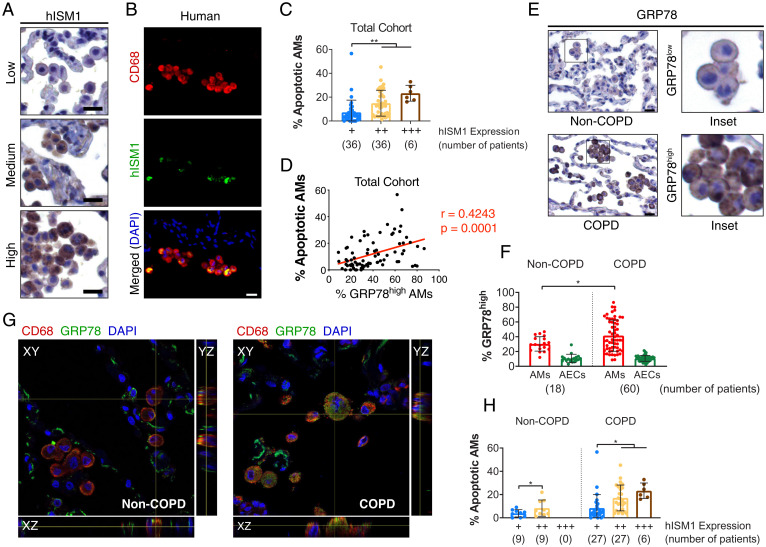
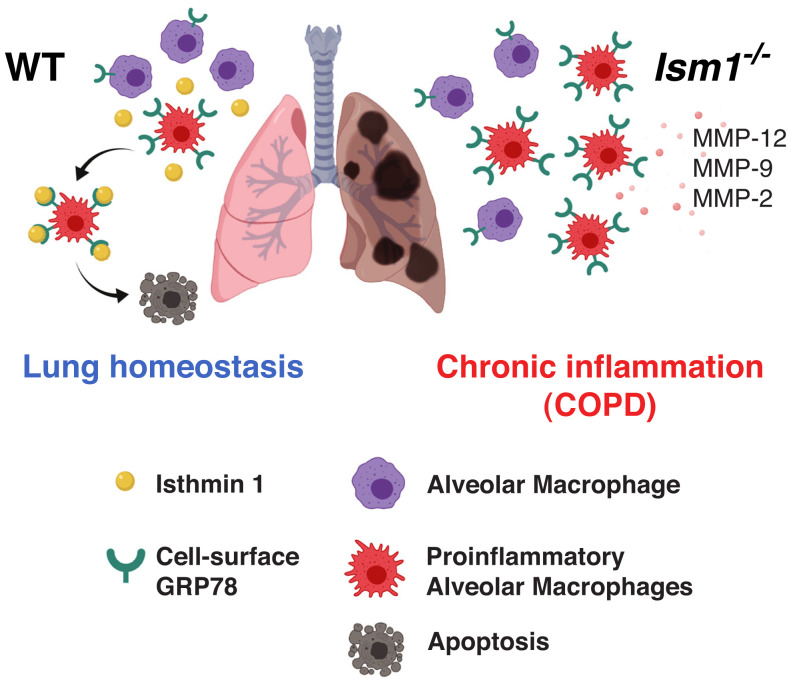
Alveolar macrophages (AMs) are critical for lung immune defense and homeostasis. They are orchestrators of chronic obstructive pulmonary disease (COPD), with their number significantly increased and functions altered in COPD. However, it is unclear how AM number and function are controlled in a healthy lung and if changes in AMs without environmental assault are sufficient to trigger lung inflammation and COPD. We report here that absence of isthmin 1 (ISM1) in mice (Ism1-/- ) leads to increase in both AM number and functional heterogeneity, with enduring lung inflammation, progressive emphysema, and significant lung function decline, phenotypes similar to human COPD. We reveal that ISM1 is a lung resident anti-inflammatory protein that selectively triggers the apoptosis of AMs that harbor high levels of its receptor cell-surface GRP78 (csGRP78). csGRP78 is present at a heterogeneous level in the AMs of a healthy lung, but csGRP78high AMs are expanded in Ism1-/- mice, cigarette smoke (CS)-induced COPD mice, and human COPD lung, making these cells the prime targets of ISM1-mediated apoptosis. We show that csGRP78high AMs mostly express MMP-12, hence proinflammatory. Intratracheal delivery of recombinant ISM1 (rISM1) depleted csGRP78high AMs in both Ism1-/- and CS-induced COPD mice, blocked emphysema development, and preserved lung function. Consistently, ISM1 expression in human lungs positively correlates with AM apoptosis, suggesting similar function of ISM1-csGRP78 in human lungs. Our findings reveal that AM apoptosis regulation is an important physiological mechanism for maintaining lung homeostasis and demonstrate the potential of pulmonary-delivered rISM1 to target csGRP78 as a therapeutic strategy for COPD.

Keywords: ISM1; alveolar macrophages; apoptosis; cell surface GRP78; chronic obstructive pulmonary disease.


Conflict of interest statement

Competing interest statement: R.G. is the scientific founder of NovoBreeze Therapeutics Co. Ltd, a private biopharma company.

Figures

Fig. 1.
Fig. 1.
Loss of ISM1 leads to pulmonary emphysema in mice. (A) Hematoxylin and eosin (H&E)  stained left lungs and (B) mean linear intercepts (MLI) of FVB/NTac WT and Ism1−/− mice at 1, 2, 6, and 9 mo of age. (C) Whole-mount stereoscopic elastin/collagen-labeled left lungs of FVB/NTac WT and Ism1−/− mice at 6 mo of age. (D) H&E stained left lung of FVB/NTac Ism1+/− mice at 9 mo of age. (E) MLI of FVB/NTac WT, Ism1+/−, and Ism1−/− mice at 9 mo of age. (F) ISM1 protein level in FVB/NTac WT, Ism1+/−, and Ism1−/− lungs at 2 mo of age determined by enzyme-linked immunosorbent assay. (G) Pathology grading of emphysema in FVB/NTac WT, Ism1+/−, and Ism1−/− mice at 2 mo of age. (H–O) Spirometry of FVB/NTac WT and Ism1−/− mice at 2 mo of age. (H) Total lung capacity, (I) functional residual capacity, (J) residual volume, (K) static compliance, (L) dynamic compliance, (M) forced expiratory volume at 100 ms (FEV100), (N) Tiffeneau–Pinelli index (FEV100/FVC), and (O) airway resistance. Data are mean ± SD and were analyzed by two-group, two-tailed Student’s t test (B and HO), and one-way ANOVA with Tukey’s post hoc test (E and F). *P < 0.05, **P < 0.01, ***P < 0.001, and ****P < 0.0001. n = 3 to 7 mice per group. (Scale bars, 200 μm for A, C, and D.) Data from A and B are integrated from two independent experiments (biological repeats, n = 7) except for MLI data for 1- and 6-mo-old mouse groups, which are from one independent experiment (n = 4). Data from CG are independent experiments using different WT and Ism1−/− mice. Data from HO are independent experiments using the same WT and Ism1−/− mice.
Fig. 2.
Fig. 2.
Ism1−/− mouse lungs present up-regulated COPD mediators at 2 mo of age. (A) H&E stained lungs showing focal AM accumulation in FVB/NTac Ism1−/− mice. (B) Liu-stained cytospins and (C) quantifications of BALF cells from FVB/NTac WT and Ism1−/− lungs. (D) Flow cytometric analysis and quantifications of AMs (Siglec F+ CD11c+) in BALF from FVB/NTac WT and Ism1−/− lungs. (E) Western blots and quantification of fold changes for Pro-MMP-12, Active-MMP-12, Pro-MMP-9, and NF-κB p65 with β-actin as loading control in FVB/NTac WT and Ism1−/− lungs. (F) IHC and quantifications of MMP-12+ and MMP-9+ AMs of FVB/NTac WT and Ism1−/− lungs. (G) IF staining for NF-κB p65 with nuclei counterstain (DAPI) and quantification of primary AMs harboring nuclear p65+ from FVB/NTac WT and Ism1−/− mice. (H) Western blot and fold change for GM–CSF with β-actin as loading control in FVB/NTac WT and Ism1−/− lungs. (I) IHC for ISM1, and (J) IHC for GRP78 and quantification of GRP78high AMs in FVB/NTac WT and Ism1−/− mice. (K) Confocal fluorescent microscopy image demonstrating csGRP78high AMs in FVB/NTac WT and Ism1−/− lungs. (L) rISM1 induces apoptosis in WT primary AMs. (M) Apoptosis of freshly isolated primary AMs from FVB/NTac WT and Ism1−/− lungs. Analysis was carried out in triplicate or quadruplicate wells. (N) Proliferation of primary AMs from WT and Ism1−/− mice. Analysis was carried out in triplicate wells. Data are mean ± SD and were analyzed by two-group, two-tailed Student’s t test (CH, J, and LN). *P < 0.05, **P < 0.01, ***P < 0.001, and ****P < 0.0001. n = 3 to 10 mice per group. A.U.: arbitrary units. (Scale bars, 20 μm for A, B, F, G, I, and J.) Data from C and D are integrated from two independent experiments using different WT and Ism1−/− mice (biological repeats, n = 9 to 10). Data from EM are representatives of twice-repeated experiments with similar results. Data from N is from one independent experiment.
Fig. 3.
Fig. 3.
Intratracheal-delivered rISM1 alleviates emphysema in Ism1−/− and CS-exposed mice. (A) Quantification of AMs from Ism1−/− lung under PBS, rISM1, and liposome-clodronate (CLO) treatments. (B) IF staining and quantifications for GRP78highCD68+ AMs, nuclei are stained by DAPI. (C) Representative H&E-stained lungs of 2-mo-old FVB/NTac Ism1−/− mice after vehicle (PBS), 1 μg rISM1, 5 μg rISM1, and CLO treatments. (D) Quantifications of MLI and (E) FEV100/FVC of untreated and treated mice groups in A. (F) Experimental design of 2-wk and (G) 8-wk CS-induced COPD model in WT BALB/cAnNTac (WT BALB/c) mice. Room air–exposed WT BALB/c mice (Sham) and CS-exposed WT BALB/c mice (CS) with vehicle (PBS) or rISM1 (10 µg rISM1) treatments at frequency and intervals indicated. (H) Quantifications of BALF cells from experimental groups in F. (I) H&E stained lungs of experimental groups in (G) depicting immune cell infiltration. n = 5 mice per group. (J) Confocal fluorescent microscopy demonstrating cell-surface expression of GRP78 in AMs in Sham and CS mouse lungs in G. (K) IF staining and quantifications of GRP78high AMs and (L) total AMs for experiment groups in G. (M) Active MMP-12 detected by Western blot and their quantifications for experiment groups in G. (N) MLI and (O) FEV100/FVC of experimental groups in G. Data are mean ± SD and were analyzed by two-group, two-tailed Student’s t test (B, D, and E: WT and Ism1−/− comparisons; and K) and one-way ANOVA with Tukey’s post hoc test (A, D, E, H, and LO). *P < 0.05, **P < 0.01, ***P < 0.001, and ****P < 0.0001, #: no significant difference compared to Sham group. n = 3 to 5 mice per group. (Scale bars: 20 μm for B and K, 50 μm for C and I.) Data from AE are representatives of twice-repeated experiments with similar results. Data from H is representative of one independent experiment. Data from I–O are independent experiments using the same experimental groups of mice in L.
Fig. 4.
Fig. 4.
hISM1 expression correlates with AM apoptosis. (A) IHC staining for hISM1 and (B) double IF staining for CD68 and hISM1 with nuclei (DAPI) counterstain in lung tissue sections showing AM expression of hISM1. (C) Quantifications of AM apoptosis in different hISM1 expression level groups. (D) Correlation analyses between high GRP78 expression with AM apoptosis. (E) Representative IHC for GRP78 depicting low GRP78 expression in non-COPD AMs and high GRP78 expression in COPD AMs. (F) Percentage of AMs and alveolar epithelial cells (AECs) in non-COPD and COPD patients with GRP78high expression (E, COPD Inset). (G) Confocal microscopy images for CD68, GRP78, and nuclei (DAPI) in non-COPD and COPD human lungs. (H) Percentage of apoptotic AMs stratified by COPD status and hISM1 expression. Data are mean ± SD and were analyzed by Pearson correlation (D), one-way ANOVA (C), and two-way ANOVA (F and H) with Tukey’s post hoc test. Patient sample sizes are depicted on graphs. (Scale bars, 20 μm for A, B, and E.)
Fig. 5.
Fig. 5.
Proposed mechanism of action for ISM1 in regulating AM apoptosis and lung homeostasis. (Left) Autocrine/paracrine ISM1 specifically targets and removes proinflammatory csGRP78high AMs via apoptosis to maintain lung homeostasis. (Right) Absence of ISM1 in Ism1−/− mice results in diminished apoptosis and accumulation of proinflammatory csGRP78high AMs, leading to proteinases overproduction, emphysema, and lung function decline.

References

    1. Gershon A. S., Warner L., Cascagnette P., Victor J. C., To T., Lifetime risk of developing chronic obstructive pulmonary disease: A longitudinal population study. Lancet 378, 991–996 (2011). - PubMed
    1. GBD 2015 Mortality and Causes of Death Collaborators, Global, regional, and national life expectancy, all-cause mortality, and cause-specific mortality for 249 causes of death, 1980-2015: A systematic analysis for the Global Burden of Disease Study 2015. Lancet 388, 1459–1544 (2016). - PMC - PubMed
    1. Singh D., et al. , Global strategy for the diagnosis, management, and prevention of chronic obstructive lung disease: The GOLD science committee report 2019. Eur. Respir. J. 53, 1900164 (2019). - PubMed
    1. Vogelmeier C. F., et al. , Global strategy for the diagnosis, management, and prevention of chronic obstructive lung disease 2017 report: GOLD executive summary. Eur. Respir. J. 49, 1700214 (2017). - PubMed
    1. Finkelstein R., Fraser R. S., Ghezzo H., Cosio M. G., Alveolar inflammation and its relation to emphysema in smokers. Am. J. Respir. Crit. Care Med. 152, 1666–1672 (1995). - PubMed

Publication types

MeSH terms

Substances

LinkOut - more resources