. 2022 Jan 25;119(4):e2116550119.
doi: 10.1073/pnas.2116550119.

Structure and function of an effector domain in antiviral factors and tumor suppressors SAMD9 and SAMD9L

Affiliations

Structure and function of an effector domain in antiviral factors and tumor suppressors SAMD9 and SAMD9L

Shuxia Peng et al. Proc Natl Acad Sci U S A. .

Abstract

SAMD9 and SAMD9L (SAMD9/9L) are antiviral factors and tumor suppressors, playing a critical role in innate immune defense against poxviruses and the development of myeloid tumors. SAMD9/9L mutations with a gain-of-function (GoF) in inhibiting cell growth cause multisystem developmental disorders including many pediatric myelodysplastic syndromes. Predicted to be multidomain proteins with an architecture like that of the NOD-like receptors, SAMD9/9L molecular functions and domain structures are largely unknown. Here, we identified a SAMD9/9L effector domain that functions by binding to double-stranded nucleic acids (dsNA) and determined the crystal structure of the domain in complex with DNA. Aided with precise mutations that differentially perturb dsNA binding, we demonstrated that the antiviral and antiproliferative functions of the wild-type and GoF SAMD9/9L variants rely on dsNA binding by the effector domain. Furthermore, we showed that GoF variants inhibit global protein synthesis, reduce translation elongation, and induce proteotoxic stress response, which all require dsNA binding by the effector domain. The identification of the structure and function of a SAMD9/9L effector domain provides a therapeutic target for SAMD9/9L-associated human diseases.

Keywords: NOD; innate immunity; myelodysplasia syndrome; myeloid malignancies; poxvirus.


Conflict of interest statement

The authors declare no competing interest.

Figures

Fig. 1.
Fig. 1.
The structure and molecular basis of DNA binding by SAMD9156-385. (A and B) Perpendicular views of the crystal structure of SAMD9156-385 dimer (green and cyan) in complex with a 22-nt dsDNA (yellow). (C) Secondary structures of SAMD9156-385 are labeled and shown in rainbow color from blue to red starting from the N terminus. (D) SAMD9156-385 primarily contacts the DNA backbone. Contacting residues (K198, K214, R221, K242, and K350) and a conserved noncontacting residue (K257) are shown as sticks and colored in red. The salt bridges are indicated as red dashed lines. (E) Purified SAMD9134-385 proteins (WT or with specific mutations) were incubated with 5′ 6-FAM–labeled 22-nt dsDNA (indicated with red *) at 2:1 molar ratio and ran on an agarose gel. Florescence image of the gel is shown with locations where labeled DNA migrated are indicated. Relative intensities of the complex bands normalized to the WT level are shown with SD from three replicates. (F) 5′ 6-FAM–labeled 22-nt dsDNA was mixed with increasing concentrations of the proteins, and the change in fluorescence millipolarization (ΔmP) in response to protein concentrations is shown (n = 3). Binding affinities are derived from nonlinear fit of the data. Those for K198E and R221E could not be reliably determined (ND).
Fig. 2.
Fig. 2.
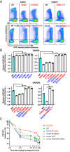
dsNA binding by DBD is essential for antiviral activities of the WT and GoF SAMD9/9L variants. (A) The predicted SAMD9/9L domain architecture and the locations of GoF (labeled in red) and DBD (labeled in blue) mutations analyzed throughout the study. SAM, sterile alpha motif; DBD, dsNA-binding domain; SIR2, silent information regulator 2; P-loop NTPase, P-loop-containing nucleoside triphosphate hydrolase; TPR, tetratricopeptide repeats; and OB, oligonucleotide/oligosaccharide-binding. (B) HEK 293T cells were transfected with mCherry-SAMD9/9L fusions for 36 h and infected with vK1C7/GFP+ for 15 h. Infection rates (GFP+%) and replication levels (GFP MFI) among SAMD9/9L-expressing and nontransfected control cells from the same culture well were simultaneously determined with flow cytometry. (C) Representative flowcytometry plots with percentage of cells in each quadrant shown. (D) Representative GFP histograms of mCherry-SAMD9+GFP+ and mCherryGFP+ cell populations with the GFP MFI shown. Relative infection rates (E) and replication levels (F) between SAMD9/9L-expressing and nontransfected cells are derived from the flow cytometry data shown in both Fig. 2 and SI Appendix, Fig. S5. Each biological replicate and SD are shown. Statistics: one-way ANOVA compared to the WT or R221E (ns, not significant; ****P <  0.0001).
Fig. 3.
Fig. 3.
dsNA binding by DBD is essential for antiproliferative activities of the WT and GoF SAMD9/9L variants. mCherry-SAMD9/9L mutants as illustrated in Fig. 2A were transfected into HEK 293T cells for 24 h, and the total and newly synthesized cellular DNA were labeled with FxCycle and EdU (for 2 h), respectively. Representative flow cytometry plots of cellular EdU and mCherry levels in the total cell population (A), or cellular EdU and FxCycle levels among mCherry-SAMD9L+ or mCherry population. (B) Relative S-phase (C, Upper) and DNA synthesis levels (C, Lower) between SAMD9-expressing and nontransfected cells from the same culture wells are derived from the flow cytometry data shown in Fig. 3 and SI Appendix, Fig. S6. Each biological replicate and SD are shown. Statistics: one-way ANOVA compared to the WT (ns, not significant; **P <  0.01; ****P <  0.0001).
Fig. 4.
Fig. 4.
GoF SAMD9 R1293W variant inhibits global protein synthesis and induces proteotoxic stress responses. BT20 cell lines that stably express either the WT or R1293W SAMD9 under the control of a doxycycline (Dox) inducible promoter were established. (A) After the cells were induced with Dox for the indicated hours, nascent cellular protein synthesis rate was measured by metabolic labeling with L-azidohomoalanine (AHA) for 2 h. SAMD9 expression is indicated by immunoblot against the Flag epitope tag, while similar loading levels are indicated by immunoblot against HSP70. (B) Transcriptome analysis of BT20 cells that were uninduced or induced to express SAMD9R1293W for 24 h. Upper: a volcano plot showing genes with significantly up- (red) and down- (blue) regulated mRNA levels upon expression of SAMD9R1293W. Genes with no significant change were shown as gray. Lower: a dot plot showing enriched pathways of significantly up-regulated genes. Enrichment of each pathway was coded by color and size of circles.
Fig. 5.
Fig. 5.
dsNA binding by DBD is essential for SAMD9/9L to inhibit global protein synthesis and reduce translation elongation. (A) HEK 293T cells were transfected with mCherry-SAMD9/9L mutants as illustrated in Fig. 2A and labeled with OPP for 30 min. Representative flow cytometry plots of OPP level relative to cellular mCherry level are shown. Background level of OPP staining in cells that were treated with cycloheximide (CHX) is shown as a control. (B) Nascent protein synthesis levels in SAMD9/9L-expressing cells relative to nontransfected cells from the same culture wells are derived from the flow cytometry data shown in Fig. 5 and SI Appendix, Fig. S8. Each replicate and SD are shown. Statistics: one-way ANOVA compared to the WT (****P <  0.0001). (C) Identically transfected cells were treated with harringtonine to block new translation initiation. Active protein synthesis level at the indicated times after harringtonine treatment was assessed by measuring OPP level as described in (A) and normalized to the level at time 0 (T0). Each data point represents the average and SD of duplicate samples.

References

    1. Narumi S., et al. , SAMD9 mutations cause a novel multisystem disorder, MIRAGE syndrome, and are associated with loss of chromosome 7. Nat. Genet. 48, 792–797 (2016). - PubMed
    1. Chen D. H., et al. , Ataxia-pancytopenia syndrome is caused by missense mutations in SAMD9L. Am. J. Hum. Genet. 98, 1146–1158 (2016). - PMC - PubMed
    1. Tesi B., et al. , Gain-of-function SAMD9L mutations cause a syndrome of cytopenia, immunodeficiency, MDS, and neurological symptoms. Blood 129, 2266–2279 (2017). - PMC - PubMed
    1. Schwartz J. R., et al. , The genomic landscape of pediatric myelodysplastic syndromes. Nat. Commun. 8, 1557 (2017). - PMC - PubMed
    1. de Jesus A. A., et al. , Distinct interferon signatures and cytokine patterns define additional systemic autoinflammatory diseases. J. Clin. Invest. 130, 1669–1682 (2020). - PMC - PubMed

Publication types

MeSH terms

Substances