. 2022 Jan 25;119(4):e2120968119.
doi: 10.1073/pnas.2120968119.

Injectable amnion hydrogel-mediated delivery of adipose-derived stem cells for osteoarthritis treatment

Affiliations

Injectable amnion hydrogel-mediated delivery of adipose-derived stem cells for osteoarthritis treatment

Maumita Bhattacharjee et al. Proc Natl Acad Sci U S A. .

Abstract

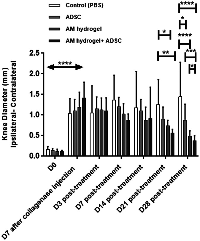
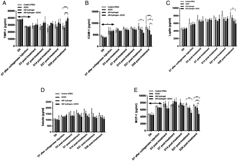
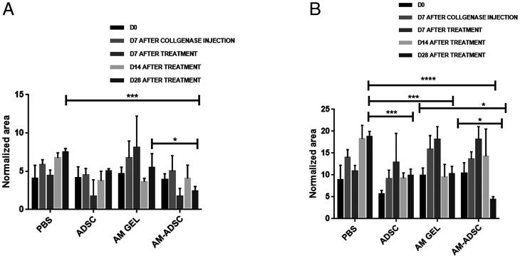
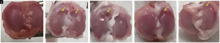
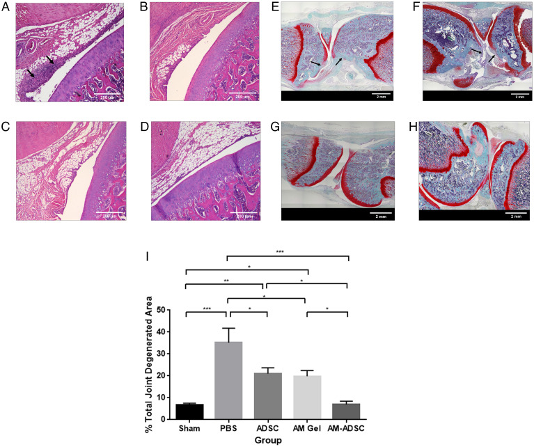
Current treatment strategies for osteoarthritis (OA) predominantly address symptoms with limited disease-modifying potential. There is a growing interest in the use of adipose-derived stem cells (ADSCs) for OA treatment and developing biomimetic injectable hydrogels as cell delivery systems. Biomimetic injectable hydrogels can simulate the native tissue microenvironment by providing appropriate biological and chemical cues for tissue regeneration. A biomimetic injectable hydrogel using amnion membrane (AM) was developed which can self-assemble in situ and retain the stem cells at the target site. In the present study, we evaluated the efficacy of intraarticular injections of AM hydrogels with and without ADSCs in reducing inflammation and cartilage degeneration in a collagenase-induced OA rat model. A week after the induction of OA, rats were treated with control (phosphate-buffered saline), ADSCs, AM gel, and AM-ADSCs. Inflammation and cartilage regeneration was evaluated by joint swelling, analysis of serum by cytokine profiling and Raman spectroscopy, gross appearance, and histology. Both AM and ADSC possess antiinflammatory and chondroprotective properties to target the sites of inflammation in an osteoarthritic joint, thereby reducing the inflammation-mediated damage to the articular cartilage. The present study demonstrated the potential of AM hydrogel to foster cartilage tissue regeneration, a comparable regenerative effect of AM hydrogel and ADSCs, and the synergistic antiinflammatory and chondroprotective effects of AM and ADSC to regenerate cartilage tissue in a rat OA model.

Keywords: adipose-derived stem cells; injectable amnion hydrogel; osteoarthritis; self-targeting property; stem cell therapy.


Conflict of interest statement

Competing interest statement: A patent titled “Injectable Amnion Hydrogel as a Cell Delivery System” has been filed and published on behalf of the inventors, C.T.L., L.S.N., and M.B. L.S.N. has competing financial interest with Soft Tissue Regeneration/Biorez. C.T.L. has the following competing financial interests: Mimedx (a company that makes amnion-based biologics), Alkermes Company, Biobind, Soft Tissue Regeneration/Biorez, and Healing Orthopaedic Technologies-Bone.

Figures

Fig. 1.
Fig. 1.
Relative abundance of proteins identified in the AM. Pie chart representing the distribution of all identified proteins in the AM according to their subcellular location (A) and function (B). Assignments were made according to their primary location and function as reported in the Human Protein Reference (http://www.hprd.org/) and UniProt (https://www.uniprot.org/) databases. Primary subcellular location: CP, cytoplasm and cytosol; CS, cytoskeleton; EC, extracellular environment; ER, endoplasmic reticulum; G, Golgi apparatus; M, mitochondrion; MB, integral to membrane and plasma membrane; N, nucleus or nucleolus; R, ribosome; V, vesicles including cytoplasmic vesicle, endosome, and lysosome. Function: EC, ECM structural constituent; I, immune response; M, metabolism and energy pathways; RN, regulation of nucleotide; SC, structural constituent of cytoskeleton; SM, structural molecule activity; ST, signal transduction; T, transporter activity; O, other functions including apoptosis, cell cycle, cell growth, motor activity, organization, extracellular ligands, and unknown. The value was rounded off to one decimal place. (C) Top five GO annotations for biological processes of the detected proteins in AM gel.
Fig. 2.
Fig. 2.
H&E staining images of rat knee joint: (A) Sham group with saline injection, (B) OA group treated with collagenase enzyme. Safranin-O staining images of rat knee joint: (C) Sham group with saline injection, (D) OA group treated with collagenase enzyme. (E) Joint swelling evaluation of sham and OA groups. n  =  6 showing mean and SD (****P < 0.0001). (Scale bars: A and B, 200 µm; C and D, 2 mm.)
Fig. 3.
Fig. 3.
Joint inflammation for different treatment groups (n = 6) showing mean and SD (****P < 0.0001, ***P < 0.001, **P < 0.01, *P < 0.05). All treatments groups showed significant decrease in joint swelling 28 d posttreatment compared to control group. The combination group (AM-ADSC) showed significant decrease in joint swelling compared to ADSCs alone and AM gel alone.
Fig. 4.
Fig. 4.
Cytokine profiling of serum isolated from whole blood from different groups: (A) TIMP-1, (B) ICAM-1, (C) leptin, (D) selectin, and (E) MCP-1; n = 6 showing mean and SD (****P < 0.0001, ***P < 0.001, **P < 0.01, *P < 0.05).
Fig. 5.
Fig. 5.
Normalized peak area of (A) region 1 (B) region 2; mean and SD with n  =  6 (****P < 0.0001, ***P < 0.001, *P < 0.05).
Fig. 6.
Fig. 6.
Gross appearance of cartilage surface after 4 wk posttreatment showing smooth surface in (A) sham group, erosion in (B) control group and (C) ADSC group, less erosion in (D) AM group, and fewer signs of lesion in (E) AM-ADSC group with closer appearance to sham group. Yellow arrows indicate the cartilage damage.
Fig. 7.
Fig. 7.
H&E staining images of rat knee joints treated with (A) control, (B) ADSC, (C) AM hydrogel, and (D) AM-ADSC after 4 wk. Safranin O staining of rat knee joint treated with (E) control, (F) ADSC, (G) AM gel, and (H) AM-ADSC. (I) Percentage joint degenerated area calculation. n = 6 showing mean and SD (***P < 0.001, **P < 0.01, *P < 0.05). (Scale bars: AD, 200 µm; EH, 2 mm.)

References

    1. Glyn-Jones S., et al. , Osteoarthritis. Lancet 386, 376–387 (2015). - PubMed
    1. Kotlarz H., Gunnarsson C. L., Fang H., Rizzo J. A., Insurer and out-of-pocket costs of osteoarthritis in the US: Evidence from national survey data. Arthritis Rheum. 60, 3546–3553 (2009). - PubMed
    1. Escobar J. L., Bhattacharjee M., Kuyinu E., Nair L. S., Laurencin C. T., Regenerative engineering for knee osteoarthritis treatment: Biomaterials and cell-based technologies. Engineering 3, 16–27 (2017). - PMC - PubMed
    1. Murab S., et al. , Matrix-embedded cytokines to simulate osteoarthritis-like cartilage microenvironments. Tissue Eng. Part A 19, 1733–1753 (2013). - PMC - PubMed
    1. Pelletier J. P., Martel-Pelletier J., Abramson S. B., Osteoarthritis, an inflammatory disease: Potential implication for the selection of new therapeutic targets. Arthritis Rheum. 44, 1237–1247 (2001). - PubMed

Publication types

MeSH terms