. 2022 Jun;126(10):1432-1438.
doi: 10.1038/s41416-022-01706-9. Epub 2022 Jan 19.

Urine DNA biomarkers for hepatocellular carcinoma screening

Affiliations

Urine DNA biomarkers for hepatocellular carcinoma screening

Amy K Kim et al. Br J Cancer. 2022 Jun.

Abstract

Background: Hepatocellular carcinoma (HCC) occurs in a well-defined high-risk patient population, but better screening tests are needed to improve sensitivity and efficacy. Therefore, we investigated the use of urine circulating tumour DNA (ctDNA) as a screening test.

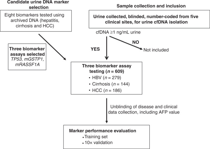
Methods: Candidate markers in urine were selected from HCC and controls. We then enrolled 609 patients from five medical centres to test the selected urine panel. A two-stage model was developed to combine AFP and urine panel as a screening test.

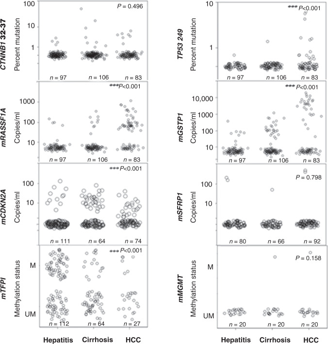
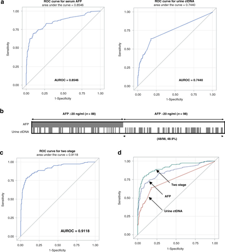
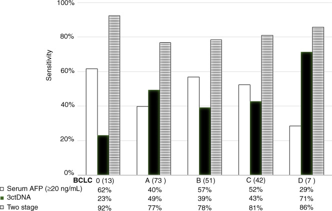
Results: Mutated TP53, and methylated RASSF1a, and GSTP1 were selected as the urine panel markers. Serum AFP outperformed the urine panel among all cases of HCC, but the urine panel identified 49% of HCC cases with low AFP < 20 ng/ml. Using the two-stage model, the combined AFP and urine panel identified 148 of the 186 HCC cases (79.6% sensitivity at 90% specificity), which was 30% more than the cases detected with serum AFP alone. It also increased early-stage HCC detection from 62% to 92% (BCLC stage 0), and 40% to 77% (BCLC stage A).

Conclusion: Urine ctDNA has promising diagnostic utility in patients in HCC, especially in those with low AFP and can be used as a potential non-invasive HCC screening test.


Conflict of interest statement

AK is a consultant to AstraZeneca, WS, SJ and SL are shareholders of JBS Science Inc. YHS has received funding from JBS Science, Inc. The remaining authors declare no competing interests.

Figures

Fig. 1
Fig. 1
Flow diagram showing outline of the study.
Fig. 2
Fig. 2. Detection of HCC-associated DNA markers in urine from patients with HCC and controls (hepatitis and cirrhosis).
The distribution of each biomarker is shown in a scatter plots by disease group and evaluated using the non-parametric independent samples Wilcoxon rank-sum test comparing HCC versus non-HCC (hepatitis and cirrhosis). The number of patient samples analysed per marker per disease category is indicated in each panel. M and UM represent qualitative measurements of methylated and unmethylated DNA.
Fig. 3
Fig. 3. Performance of urine ctDNA markers for distinguishing HCC from non-HCC.
a Receiver-operating curves (ROC) of serum AFP alone and urine ctDNA markers alone. b Distribution of patients stratified by AFP cut-off of 20 ng/mL. The marker values are summarised in Supplemental Table 5. Each box represents a patient sample, and those with positive urine ctDNA biomarker detection are filled, based on the cut-off set at 90% specificity. AFP was positive (≥20 ng/mL) in 47.3% (88/186) of all HCC cases. Urine ctDNA panel was positive in 44.1% (82/186) of all HCC cases which included 48.9% (48/98) of the low AFP (<20 ng/mL) HCC group. c ROC of the two-stage model. d Comparison of three ROC curves as indicated.
Fig. 4
Fig. 4. Sensitivities of AFP ≥ 20 ng/ml, urine ctDNA and the two-stage model in different HCC stages per BCLC criteria.
All tests were analysed at 90% specificity cut-off. The urine panel consistently selected additional HCC cases with low AFP, especially in early-stage BCLC stages 0 and A. BCLC Barcelona Clinic Liver Cancer staging. Patient numbers are shown in parenthesis in each stage.

References

    1. Davila JA, Morgan RO, Richardson PA, Du XL, McGlynn KA, El-Serag HB. Use of surveillance for hepatocellular carcinoma among patients with cirrhosis in the United States. Hepatology. 2010;52:132–41. doi: 10.1002/hep.23615. - DOI - PMC - PubMed
    1. Heimbach JK, Kulik LM, Finn RS, Sirlin CB, Abecassis MM, Roberts LR, et al. AASLD guidelines for the treatment of hepatocellular carcinoma. Hepatology. 2018;67:358–80.. doi: 10.1002/hep.29086. - DOI - PubMed
    1. Howlader N, Noone AM, Krapcho M, Miller D, Bishop K, Altekruse SF, et al. SEER cancer statistics review, 1975–2013, based on November 2015 SEER data submission, posted to the SEER website. Bethesda, MD, USA: National Cancer Institute; 2016.
    1. Ferlay JSI, Ervik M, Dikshit R, Eser S, Mathers C, Rebelo M, et al. Cancer incidence and mortality worldwide: sources, methods and major patterns in GLOBOCAN 2012. Int J Cancer. 2015;136:E359–86. doi: 10.1002/ijc.29210. - DOI - PubMed
    1. Daniele B, Bencivenga A, Megna AS, Tinessa V. α-fetoprotein and ultrasonography screening for hepatocellular carcinoma. Gastroenterology. 2004;127:S108–S12.. doi: 10.1053/j.gastro.2004.09.023. - DOI - PubMed

Publication types

MeSH terms