. 2022 Jan 11:16:93-100.
doi: 10.2147/OPTH.S344523. eCollection 2022.

Intrascleral Intraocular Lens Fixation Preserving the Lens Capsule in Cases of Cataract with Insufficient Zonular Support

Affiliations

Intrascleral Intraocular Lens Fixation Preserving the Lens Capsule in Cases of Cataract with Insufficient Zonular Support

Mutsuko Kato et al. Clin Ophthalmol. .

Abstract

Purpose: To report our modified simple technique for optic capture and the clinical results of intrascleral IOL fixation preserving the lens capsule, without vitrectomy, in cases of cataract with insufficient zonular support to stabilize the intraocular lens (IOL).

Patients and methods: In 37 eyes of 25 patients with phacodonesis and two or more risk factors for progressive zonular insufficiency, we inserted a CTR to support the capsule and zonules during cataract surgery and IOL fixation; an optic was inserted into the lens capsule, and a haptic was fixed in the scleral tunnel without vitrectomy. In all cases, anterior or total vitrectomy was not needed.

Results: The postoperative mean (± standard deviation) tilt and decentration of the implanted IOL did not change from 6 to 12 months (6.77 ± 3.15° to 6.33 ± 3.38° and 0.60 ± 0.30 to 0.61 ± 0.35 mm, respectively). We encountered no late IOL dislocation and no retinal complications, including retinal breaks or cystoid macular oedema, postoperatively (follow-up = 21.1 ± 5.2 months).

Conclusion: Our modified techniques preclude the need for vitrectomy. If the lens capsule can be preserved using a CTR, our modified technique can be used to stabilize IOL.

Keywords: capsular tension ring; insufficient zonular support; intrascleral IOL fixation; lens capsule; phacodonesis.


Conflict of interest statement

The authors report no conflicts of interest relevant to this work.

Figures
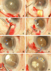
Figure 1
Figure 1
Procedure for intrascleral IOL fixation preserving the lens capsule. (A) For each case with phacodonesis, a CTR was inserted into the capsule after completion of appropriately sized CCC. PEA and I/A were completed with the aid of the CTR. (B) An IOL was implanted in the out-of-the-bag position using an injector. (C) A T-shaped 2-mm long scleral tunnel for fixation of an IOL haptic was created 2.0 mm from the limbus at the 1-o’clock Meridian (or at both the 1- and 7-o’clock meridians when the capsule support was very weak and/or the patient was relatively young). (D) Angle sclerotomy creating a T-shaped tunnel (represented by dots) was performed using a 26-G needle (arrows) parallel to the iris and above the anterior capsule. (E) The upper haptics of the IOL (arrow head) were dialled in and externalized through the sclerotomy site using 25-G forceps (arrow). (F) The tip of an IOL haptic (arrow) was inserted into the scleral tunnel (“intrascleral haptic fixation”). (G) The scleral incision was closed with a 7–0 vicryl suture; we included a haptic (arrow) to prevent intraocular fluid leakage and the IOL from shifting. (H) The IOL optic was gently inserted into the capsular bag (“optic capture”). The arrows indicate the CCC margin.
Figure 2
Figure 2
Schema of the intrascleral IOL fixation method preserving the lens capsule. (2-1) First, an IOL was implanted in an out-of-the-bag position. Then, the tip of the upper IOL haptic was fixed in the scleral T-shaped tunnel (the lower haptic was placed on the bag or fixed in the scleral tunnel, as was the upper haptic). (2-2) Second, the IOL optic was gently inserted into the capsular bag (“optic capture”). The IOL supports the capsule containing the CTR. Simultaneously, the spared capsule and vitreous body support the optic (to prevent IOL tilting). Slice “A” shows the optics of the implanted in-the-bag IOL and the positions of the spared capsule and CTR. Slice “B” shows that the haptics support the capsule with the CTR and that the optic is supported by the face of the spared capsule and vitreous, preventing tilting. (2-3) The arrowhead indicates the tip of the upper IOL haptic and 7–0 vicryl suture. (2-4) The arrows indicate the CCC margin, and the arrowhead indicates the loop attachment.

References

    1. Gimbel HV, Condon GP, Kohnen T, et al. Late in-the-bag intraocular lens dislocation: incidence, prevention, and management. J Cataract Refract Surg. 2005;31(11):2193–2204. doi:10.1016/j.jcrs.2005.06.053 - DOI - PubMed
    1. Ascaso FJ, Huerva V, Grzybowski A. Epidemiology, etiology, and prevention of late IOL-capsular bag complex dislocation: review of the literature. J Ophthalmol. 2015;2015:805706. doi:10.1155/2015/805706 - DOI - PMC - PubMed
    1. Dabrowska-Kloda K, Kloda T, Boudiaf S, et al. Incidence and risk factors of late in-the-bag intraocular lens dislocation: evaluation of 140 eyes between 1992 and 2012. J Cataract Refract Surg. 2015;41(7):1376–1382. doi:10.1016/j.jcrs.2014.10.040 - DOI - PubMed
    1. Mönestam E. Frequency of intraocular lens dislocation and pseudophacodonesis, 20 years after cataract surgery – a prospective study. Am J Ophthalmol. 2019;198:215–222. doi:10.1016/j.ajo.2018.10.020 - DOI - PubMed
    1. Lee G-I, Lim DH, Chi SA, et al. Risk factors for intraocular lens dislocation after phacoemulsification: a nationwide population-based cohort study. Am J Ophthalmol. 2020;214:86–96. doi:10.1016/j.ajo.2020.03.012 - DOI - PubMed