Huge Thoracic Aortic Aneurysm Presenting with Jaundice: A Case Report
- PMID: 35046660
- PMCID: PMC8762516
- DOI: 10.2147/VHRM.S346041
Huge Thoracic Aortic Aneurysm Presenting with Jaundice: A Case Report
Abstract
Introduction: Thoracic aortic aneurysms (TAA) are encountered frequently in the emergency department with an obscure presentation. Most of these aneurysms are incidentally discovered while doing routine imaging studies. This report describes a case of unusual presentation of TAAs.
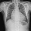
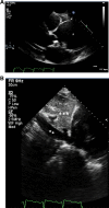
Case presentation: A 37-year-old male presented to the emergency department with jaundice, shortness of breath, abdominal swelling, and lower limb edema, which worsened during the previous month. The patient was worked up and diagnosed with an ascending aortic aneurysm measuring 6.9 cm associated with severe aortic insufficiency and heart failure.
Conclusion: TAA is a life-threatening condition with indistinct signs and symptoms. A high index of suspicion and early implementation of radiological studies are prerequisites to reach a diagnosis.
Keywords: ascending aortic aneurysm; clinical presentation; jaundice; thoracic aortic aneurysm; transthoracic echocardiography.
© 2022 Al Mulhim.
Conflict of interest statement
The author reports no conflicts of interest in this work.
Figures


References
Publication types
MeSH terms
LinkOut - more resources
Full Text Sources

