Review
. 2022 Jan 3:15:778900.
doi: 10.3389/fncel.2021.778900. eCollection 2021.

Red Light Optogenetics in Neuroscience

Affiliations
Review

Red Light Optogenetics in Neuroscience

Kimmo Lehtinen et al. Front Cell Neurosci. .

Abstract

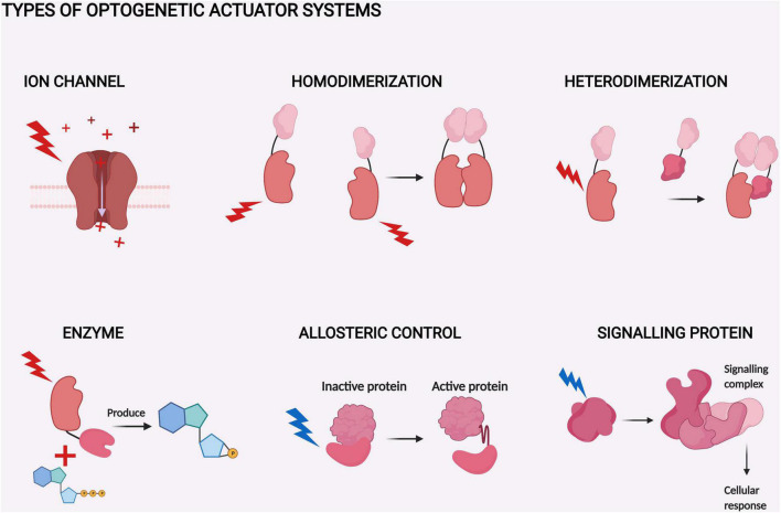
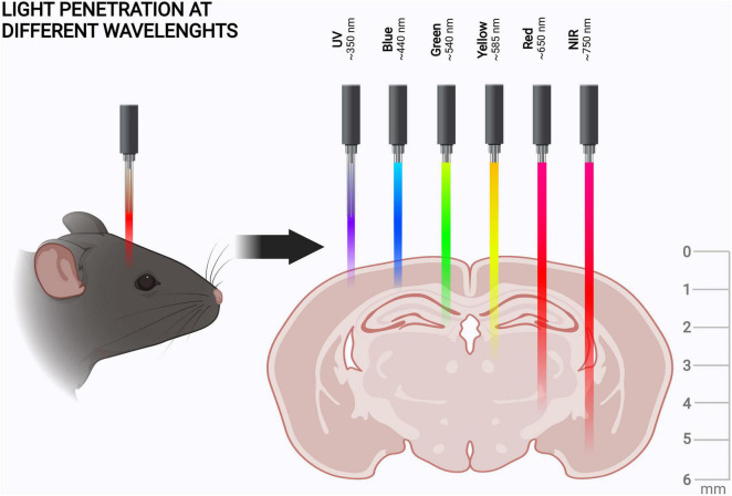
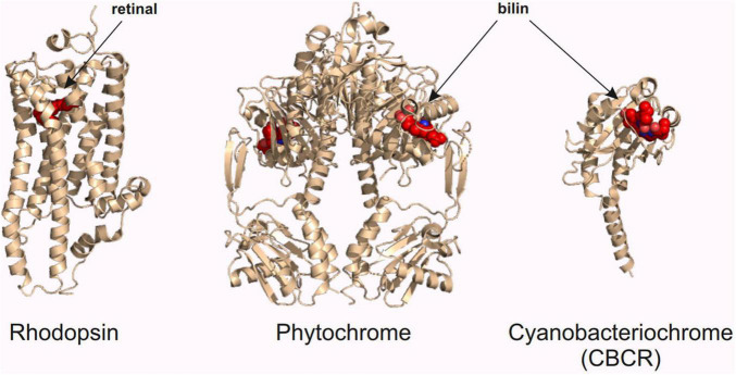
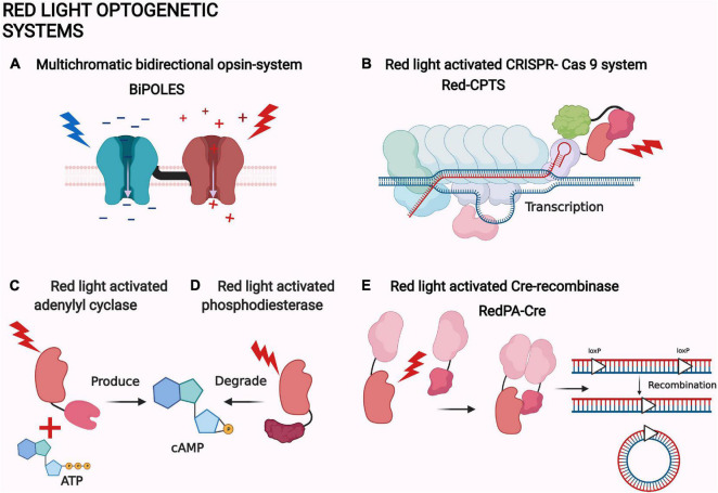
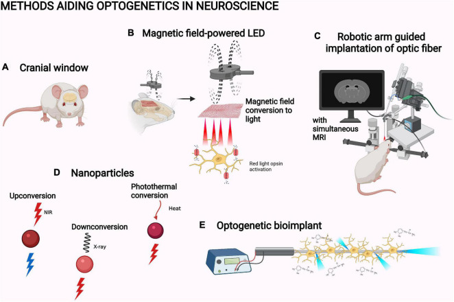
Optogenetics, a field concentrating on controlling cellular functions by means of light-activated proteins, has shown tremendous potential in neuroscience. It possesses superior spatiotemporal resolution compared to the surgical, electrical, and pharmacological methods traditionally used in studying brain function. A multitude of optogenetic tools for neuroscience have been created that, for example, enable the control of action potential generation via light-activated ion channels. Other optogenetic proteins have been used in the brain, for example, to control long-term potentiation or to ablate specific subtypes of neurons. In in vivo applications, however, the majority of optogenetic tools are operated with blue, green, or yellow light, which all have limited penetration in biological tissues compared to red light and especially infrared light. This difference is significant, especially considering the size of the rodent brain, a major research model in neuroscience. Our review will focus on the utilization of red light-operated optogenetic tools in neuroscience. We first outline the advantages of red light for in vivo studies. Then we provide a brief overview of the red light-activated optogenetic proteins and systems with a focus on new developments in the field. Finally, we will highlight different tools and applications, which further facilitate the use of red light optogenetics in neuroscience.

Keywords: brain; near-infrared; neuron; neuroscience; opsin; optogenetics; phytochrome.


Conflict of interest statement

The authors declare that the research was conducted in the absence of any commercial or financial relationships that could be construed as a potential conflict of interest.

Figures

FIGURE 1
FIGURE 1
Types of optogenetic actuator systems. The six major actuator systems and the example studies related to them pertain to ion channels (Klapoetke et al., 2014), homodimerization (Schmidl et al., 2019), heterodimerization (Kaberniuk et al., 2016), enzyme activity (Gasser et al., 2014), allosteric control (Strickland et al., 2008), and signaling proteins (Kim et al., 2005). The components of the systems with similar function are depicted with a similar shape. The shapes are not accurate depictions of the actual proteins and are not to scale. The illumination of each actuator is depicted as lightning and colored according to each system. The figure was created with BioRender.com.
FIGURE 2
FIGURE 2
Tissue penetration of different wavelengths of light. Tissue penetration is illustrated from the coronal view of the rodent brain. The light penetration depths, scales, and shown wavelengths are indicative (Jacques, 2013; Diem et al., 2016; Ash et al., 2017). The figure was created with BioRender.com.
FIGURE 3
FIGURE 3
Red light optogenetic actuator classes. Example structures of the three classes of the red light optogenetic actuators. Note that only the photosensory fragment of each protein is shown. The structures are to scale relative to each other. Each structure was derived from the following Protein Data Bank (Berman et al., 2000) coordinates: 3AYN (rhodopsin) (Murakami and Kouyama, 2011), 4O0P (phytochrome) (Takala et al., 2014), and 6UV8 (cyanobacteriochrome) (Bandara et al., 2021). The figure was created with the PyMOL Molecular Graphics System version 2.3.3 (Schrödinger, LLC).
FIGURE 4
FIGURE 4
Examples of red light optogenetic systems. (A) Multichromatic directional opsin system BiPOLES (Vierock et al., 2021). (B) Red light-activated CRISPR-Cas 9 system Red-CPTS (Kuwasaki et al., 2021). (C) Red light-activated adenylyl cyclase (Fomicheva et al., 2019). (D) Red light-activated phosphodiesterase (Stabel et al., 2019). (E) Red light-activated Cre-recombinase RedPA-Cre (Kuwasaki et al., 2021). The same protein components are depicted with a similar shape in each panel. The shapes are not accurate depictions of the proteins and are not to scale. The illumination of each actuator is depicted as lightning and colored according to each system. The figure was created with BioRender.com.
FIGURE 5
FIGURE 5
Methods aiding optogenetics in neuroscience. (A) Cranial window (Cha et al., 2020). (B) Magnetic field-powered LED (Lee et al., 2020). (C) Robotic arm guided implantation with magnetic resonance imaging, MRI (Chen et al., 2019). (D) Upconversion nanoparticle, UPNC (Wang et al., 2019), X-ray downconverting nanoparticle (Chen et al., 2021), and photothermal-converting nanoparticle (Chen X. et al., 2020; Qiao et al., 2021). (E) Optogenetic bioimplant (Vasudevan et al., 2019). The objects are not to scale. The figure was created with BioRender.com.

References

    1. Accanto N., Chen I., Ronzitti E., Molinier C., Tourain C., Papagiakoumou E., et al. (2019). Multiplexed temporally focused light shaping through a gradient index lens for precise in-depth optogenetic photostimulation. Sci. Rep. 9 1–10. 10.1038/s41598-019-43933-w - DOI - PMC - PubMed
    1. Adli M. (2018). The CRISPR tool kit for genome editing and beyond. Nat. Commun. 9 1–13. 10.1038/s41467-018-04252-2 - DOI - PMC - PubMed
    1. Al-Juboori S., Dondzillo A., Stubblefield E., Felsen G., Lei T., Klug A. (2013). Light scattering properties vary across different regions of the adult mouse brain. PLoS One 8:e67626. 10.1371/journal.pone.0067626 - DOI - PMC - PubMed
    1. Anderson R., Farinelli W., Laubach H., Manstein D., Yaroslavsky A., Gubeli J., et al. (2006). Selective photothermolysis of lipid-rich tissues: a free electron laser study. Lasers Surg. Med. 38 913–919. 10.1002/LSM.20393 - DOI - PubMed
    1. Ash C., Dubec M., Donne K., Bashford T. (2017). Effect of wavelength and beam width on penetration in light-tissue interaction using computational methods. Lasers Med. Sci. 32 1909–1918. 10.1007/s10103-017-2317-4 - DOI - PMC - PubMed

LinkOut - more resources