. 2022 Jan 10:2022:2485250.
doi: 10.1155/2022/2485250. eCollection 2022.

STAT6/VDR Axis Mitigates Lung Inflammatory Injury by Promoting Nrf2 Signaling Pathway

Affiliations

STAT6/VDR Axis Mitigates Lung Inflammatory Injury by Promoting Nrf2 Signaling Pathway

Youjing Yang et al. Oxid Med Cell Longev. .

Abstract

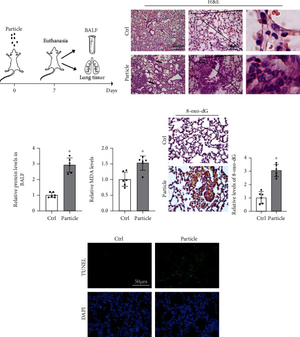
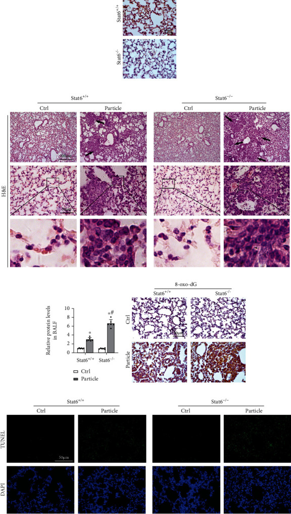
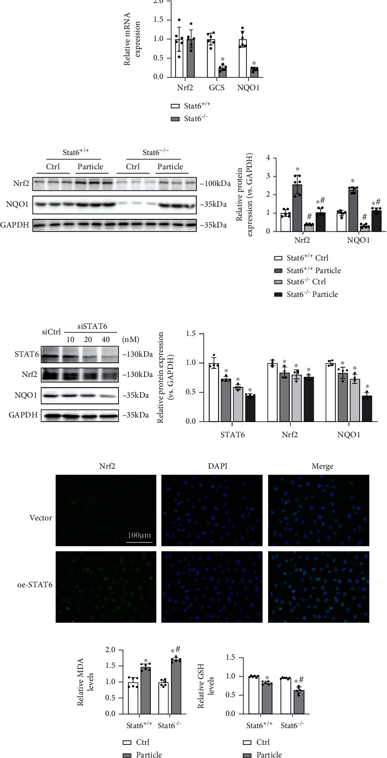
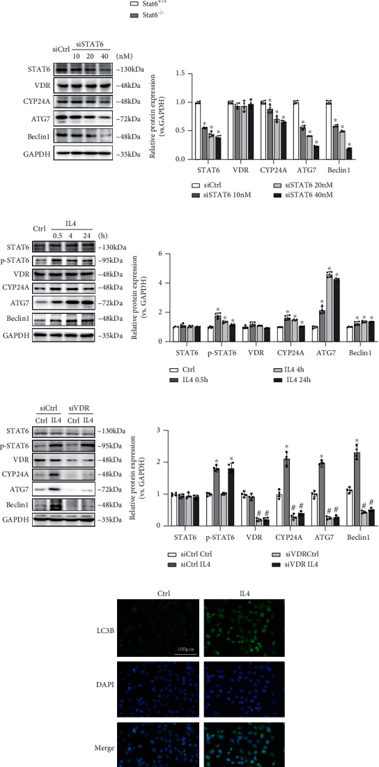
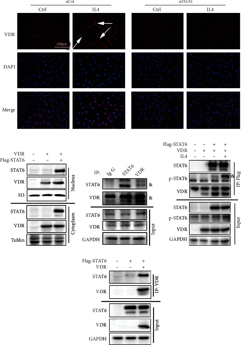
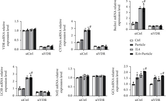
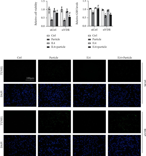
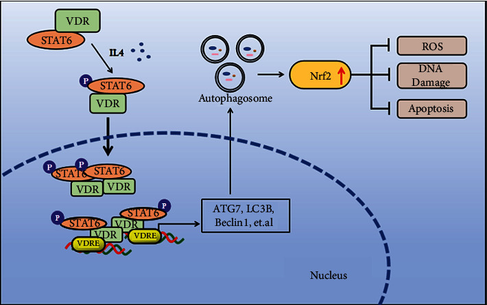
Lung inflammatory injury is a global public health concern. It is characterized by infiltration of diverse inflammatory cells and thickening of pulmonary septum along with oxidative stress to airway epithelial cells. STAT6 is a nuclear transcription factor that plays a crucial role in orchestrating the immune response, but its function in tissue inflammatory injury has not been comprehensively studied. Here, we demonstrated that STAT6 activation can protect against particle-induced lung inflammatory injury by resisting oxidative stress. Specifically, genetic ablation of STAT6 was observed to worsen particle-induced lung injury mainly by disrupting the lungs' antioxidant capacity, as reflected by the downregulation of the Nrf2 signaling pathway, an increase in malondialdehyde levels, and a decrease in glutathione levels. Vitamin D receptor (VDR) has been previously proved to positively regulate Nrf2 signals. In this study, silencing VDR expression in human bronchial epithelial BEAS-2B cells consistently suppressed autophagy-mediated activation of the Nrf2 signaling pathway, thereby aggravating particle-induced cell damage. Mechanically, STAT6 activation promoted the nuclear translocation of VDR, which increased the transcription of autophagy-related genes and induced Nrf2 signals, and silencing VDR abolished these effects. Our research provides important insights into the role of STAT6 in oxidative damage and reveals its potential underlying mechanism. This information not only deepens the appreciation of STAT6 but also opens new avenues for the discovery of therapies for inflammatory respiratory system disorders.


Conflict of interest statement

The authors have declared that no competing interest exists.

Figures

Figure 1
Figure 1
Particle caused severe oxidative injury in lung tissue. (a) Pattern of mouse model procedure. Mice received particles intratracheally at day 0 and sacrificed at 7th day. BALF and lung tissue were harvested for next analysis. (b) Representative H&E-stained lung sections (n = 6, black arrows indicated pulmonary nodule, and infiltration of inflammatory cells was magnified at right panel). (c) The protein content of BALF was measured. Results were shown as means ± SD (n = 6, P < 0.05, Ctrl vs. particle). (d) Relative values of MDA in lung tissue from each group were measured. Results were expressed as means ± SD (n = 6, P < 0.05, Ctrl vs. particle). (e) IHC staining of 8-oxo-dG in the lung tissue from the indicated group was performed and quantified. Representative images from each group were shown. Results were expressed as means ± SD (n = 6, P < 0.05, Ctrl vs. particle). (f) Representative images of TUNEL assay staining in lung tissue from each group were shown.
Figure 2
Figure 2
STAT6 deficiency exacerbated particle caused lung oxidative injury. (a) Representative photographs of lung tissue STAT6 staining from Stat6+/+ and Stat6−/− mice. (b) Representative H&E-stained lung sections (black arrows indicated pulmonary nodule, and infiltration of inflammatory cells was magnified at the below). (c) The protein content of BALF from the indicated group was measured. Results were expressed as means ± SD (n = 6, P < 0.05, Ctrl vs. particle; #P < 0.05, Stat6+/+ vs. Stat6−/−). (d) IHC staining of 8-oxo-dG in lung tissue sections from the indicated groups was performed. (e) TUNEL staining of lung tissue. Representative images from each group were shown.
Figure 3
Figure 3
Deficiency of STAT6 impaired the antioxidant capacity of lung tissues by downregulating Nrf2 signaling. (a) The mRNA expressions of Nrf2, GCS, and NQO1 in lung tissues were detected by qRT-PCR. Results were expressed as means ± SD (n = 6, P < 0.05, Stat6+/+ vs. Stat6−/−). (b) The protein expression levels of Nrf2 and NQO1 in lung tissues were determined by western blot analysis in the indicated group. Results were presented as means ± SD (n = 6, P < 0.05, Ctrl vs. particle; #P < 0.05, Stat6+/+ vs. Stat6−/−). (c) The protein expressions of STAT6, Nrf2, and NQO1 in BEAS-2B cells treated with siCtrl or indicated doses of siSTAT6 were measured using western blot analysis. Results were expressed as means ± SD (n = 4; P < 0.05, Ctrl vs. treatment groups). (d) Immunofluorescence staining of Nrf2 in the cells with transfection of STAT6 plasmid or vector. Nuclei were stained with DAPI (blue). (e) Relative MDA and (f) GSH levels in lung tissue were measured. Results were expressed as means ± SD (n = 6, P < 0.05, Ctrl vs. particle; #P < 0.05, Stat6+/+ vs. Stat6−/−).
Figure 4
Figure 4
STAT6 positively regulates autophagy through VDR. (a) The mRNA levels of STAT6, Arg1, VDR, CYP24A, ATG7, Beclin1, and LC3B in lung tissue were measured by qRT-PCR assay. Results were expressed as means ± SD (n = 6, P < 0.05, Stat6+/+ vs. Stat6−/−). (b) The protein expressions of STAT6, VDR, CYP24A, ATG7, and Beclin1 in BEAS-2B cells following different doses of siSTAT6 were measured using western blot analysis (n = 4; P < 0.05, Ctrl vs. treatment groups). (c) The protein expressions of STAT6, p-STAT6, VDR, CYP24A, ATG7, and Beclin1 in BEAS-2B cells following different doses of IL4 were measured using western blot analysis (n = 4; P < 0.05, Ctrl vs. treatment groups). (d) Cells were transfected with siVDR for 24 h and followed by IL4 (20 ng/ml, 24 h) treatment. The protein expressions of STAT6, p-STAT6, VDR, CYP24A, ATG7, and Beclin1 were detected by western blot analysis (n = 4; P < 0.05, Ctrl vs. IL4; #P < 0.05, siCtrl vs. siVDR). (e) Immunofluorescence staining of LC3B in the cells with IL4 treatment. Nuclei were stained with DAPI (blue).
Figure 5
Figure 5
The interaction of STAT6 and VDR. (a) Immunofluorescence staining of VDR in the cells with or without siSTAT6 20 nM 48 h transfection and IL4 (20 ng/ml, 24 h) treatment. Nuclei were stained with DAPI (blue). (b) The protein expression of STAT6 and VDR in the nucleus and cytoplasm after transfection of VDR or STAT6 plasmid in BEAS-2B cells. (c) The protein complex of STAT6-VDR in BEAS-2B cells was assayed by immunoprecipitation to analyze the association between STAT6 and VDR at the endogenous level (and indicate the target bind). (d) HEK293T cells overexpressed different combinations of Flag-STAT6 and VDR as indicated. Exogenous STAT6 interacted with VDR was assayed by immunoprecipitation with Flag beads. (e) HEK293T cells were overexpressed with indicated plasmids (Flag-STAT6 and VDR), and the interaction between VDR and STAT6 was detected by immunoprecipitation using agarose beads along with VDR antibodies.
Figure 6
Figure 6
Autophagy-related genes and Nrf2 signaling pathway were decreased with blockade of VDR. BEAS-2B cells were transfected with either Ctrl siRNA or VDR siRNA for 24 h in serum-free medium and then received indicated medium from THP-1 cells (Ctrl and particle groups) with or without IL4 treatment for another 24 h. The total RNA was extracted, and qRT-PCR assay was employed to measure the mRNA expression of (a) VDR, (b) CYP24A, (c) Beclin1, (d) LC3B, (e) Nrf2, and (f) GCS. Results were expressed as means ± SD (n = 4; P < 0.05, Ctrl vs. treatment groups; #P < 0.05, P vs. IL4+P).
Figure 7
Figure 7
STAT6 protects against oxidative damage through VDR. BEAS-2B cells were transfected with either Ctrl siRNA or VDR siRNA for 24 h in serum-free medium followed by particle medium exposure with or without IL4 24 h administration. (a) The viability of BEAS-2B cells was measured, and results were shown as means ± SD (n = 4; P < 0.05, Ctrl vs. particle; #P < 0.05, particle vs. IL4+particle). (b) The GSH levels in the indicated group were detected. Results are expressed as means ± SD (n = 4; P < 0.05, Ctrl vs. P; #P < 0.05, P vs. IL4+P). (c) Representative photographs of TUNEL assay staining in BEAS-2B cells from the indicated group were shown.
Figure 8
Figure 8
Proposed model for the STAT6/VDR axis mitigates lung inflammatory. STAT6 activation enhances the translocation of VDR to the nucleus, which increased the transcription of autophagy-related genes and then induced Nrf2 signaling, preventing oxidative stress, DNA damage, and apoptosis.

Similar articles

Cited by

References

    1. Lee H., Abston E., Zhang D., Rai A., Jin Y. Extracellular vesicle: an emerging mediator of intercellular crosstalk in lung inflammation and injury. Frontiers in Immunology . 2018;9:p. 924. doi: 10.3389/fimmu.2018.00924. - DOI - PMC - PubMed
    1. Zhao C., Yang X., Su E. M., et al. Signals of vagal circuits engaging with AKT1 in α7 nAChR+CD11b+ cells lessen _E. coli_ and LPS-induced acute inflammatory injury. Cell Discovery . 2017;3(1):p. 17009. doi: 10.1038/celldisc.2017.9. - DOI - PMC - PubMed
    1. Xian H., Liu Y., Rundberg Nilsson A., et al. Metformin inhibition of mitochondrial ATP and DNA synthesis abrogates NLRP3 inflammasome activation and pulmonary inflammation. Immunity . 2021;54(7):1463–1477.e11. doi: 10.1016/j.immuni.2021.05.004. - DOI - PMC - PubMed
    1. Wang M., Li J., Dong S., et al. Silica nanoparticles induce lung inflammation in mice via ROS/PARP/TRPM2 signaling-mediated lysosome impairment and autophagy dysfunction. Particle and Fibre Toxicology . 2020;17(1):p. 23. doi: 10.1186/s12989-020-00353-3. - DOI - PMC - PubMed
    1. Quezada-Maldonado E. M., Sánchez-Pérez Y., Chirino Y. I., García-Cuellar C. M. Airborne particulate matter induces oxidative damage, DNA adduct formation and alterations in DNA repair pathways. Environmental Pollution . 2021;2021(287, article 117313) - PubMed

Substances