

Falciform ligament torsion as a rare aetiology of the acute abdomen
- PMID: 35047164
- PMCID: PMC8763607
- DOI: 10.1093/jscr/rjab150
Falciform ligament torsion as a rare aetiology of the acute abdomen
Abstract
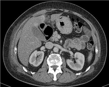
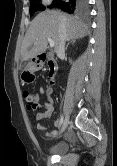
The falciform ligament is a remnant of the embryonic ventral mesentery containing the obliterated umbilical vein and round ligament. It extends from the umbilicus to the superior aspect of the diaphragm. We report about a 53-year-old fit and well patient who presented with acute upper abdominal pain with tenderness to palpation. Ultrasound scan was unremarkable, but blood tests revealed raised inflammatory markers. Thus, computed tomography was performed. This demonstrated acute torsion and fat necrosis of the falciform ligament, which was the aetiology of the upper abdominal pain. Such pathology is rare with 23 previously reported cases. Conservative management is usually proposed, but on occasion, surgical intervention may be warranted in cases that do not respond to initial supportive measures. We describe this case to demonstrate a rare cause of a common presentation to the surgical service.
Published by Oxford University Press and JSCR Publishing Ltd. All rights reserved. © The Author(s) 2022.
Figures
References
-
- Uyttenhove F, Leroy C, Nzamushe Lepan Mabla JR, Ernst O. Torsion of a fatty fringe of the falciform ligament, a rare cause of right hypochondrial pain. Diagn Interv Imaging 2013;94:637–9. - PubMed
-
- Li XP, Xu DC, Tan HY, Li CL. Anatomical study on the morphology and blood supply of the falciform ligament and its clinical significance. Surg Radiol Anat 2004;26:106–9. - PubMed
-
- Coulier B. Contribution of US and CT for diagnosis of intraperitoneal focal fat infarction (IFFI): a pictorial review. JBR-BTR 2010;93:171–85. - PubMed
-
- Lloyd T. Primary torsion of the falciform ligament: computed tomography and ultrasound findings. Australas Radiol 2006;50:252–4. - PubMed
Publication types
LinkOut - more resources
Full Text Sources
