

Magnetic resonance imaging pattern variability in dysferlinopathy
- PMID: 35047756
- PMCID: PMC8744010
- DOI: 10.36185/2532-1900-059
Magnetic resonance imaging pattern variability in dysferlinopathy
Abstract
The widespread use of magnetic resonance imaging (MRI) in the diagnosis of myopathies has made it possible to clarify the typical MRI pattern of dysferlinopathy. However, sufficient attention has not been given to the variability of MRI patterns in dysferlinopathy.
Materials and methods: Twenty-five patients with the clinical manifestations of dysferlinopathy were examined. For all patients, creatine phosphokinase levels were measured and molecular genetics were examined. In two patients, immunohistochemical examinations of muscle biopsies were performed. MRI scanning was included T2 multi-slice multi-echo, T1 weighted, T2 weighted and Short Tau Inversion Recovery T2 weighted sequences. Quantitative and semi-quantitative evaluations of fatty replacement and swelling of the muscles were undertaken.
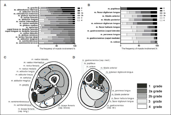
Results: Variability in the MRI patterns was lowest in the pelvis and leg muscles and highest in the thigh muscles. Three main types of MRI patterns were distinguished: posterior-dominant (80%), anterior-dominant (16%), and diffuse (4%). Among patients with the anterior-dominant pattern, the collagen-like variant (4%), proximal variant (4%) and pseudo-myositis (8%) were separately distinguished.
Conclusions: Awareness of atypical MRI patterns in dysferlinopathy is important for increasing the efficiency of routine diagnostics and optimizing the search for causative gene mutations.
Keywords: LGMD2B; LGMDR2; MRI pattern; Miyoshi myopathy; T2-MSME; dysferlinopathy.
©2021 Gaetano Conte Academy - Mediterranean Society of Myology, Naples, Italy.
Figures


References
-
- Aoki M, Liu J, Richard I, et al. . Genomic organization of the dysferlin gene and novel mutations in Miyoshi myopathy. Neurology 2001;57:271-278. https://doi.org/10.1212/wnl.57.2.271 10.1212/wnl.57.2.271 - DOI - PubMed
-
- Okahashi S, Ogawa G, Suzuki M, et al. . Asymptomatic sporadic dysferlinopathy presenting with elevation of serum creatine kinase. Typical distribution of muscle involvement shown by MRI but not by CT. Int. Mede (Tokyo, Japan) 2008;47:305-307. https://doi.org/10.2169/internalmedicine.47.0519 10.2169/internalmedicine.47.0519 - DOI - PubMed
-
- Illa I, Serrano-Munuera C, Gallardo E, et al. . Distal anterior compartment myopathy: a dysferlin mutation causing a new muscular dystrophy phenotype. Ann Neurol 2001;49:130-134. PMID: . - PubMed
-
- Paradas C, Gonzalez-Quereda L, De Luna N, et al. . A new phenotype of dysferlinopathy with congenital onset. Neuromusc Disord 2009;19:21-25. https://doi.org/10.1016/j.nmd.2008.09.015 10.1016/j.nmd.2008.09.015 - DOI - PubMed
-
- Diaz-Manera J, Fernandez-Torron R, Llauger J, et al. . Muscle MRI in patients with dysferlinopathy: pattern recognition and implications for clinical trials. J Neurol Neurosurg Psychiatry 2018;89:1071-1081. https://doi.org/10.1136/jnnp-2017-317488 10.1136/jnnp-2017-317488 - DOI - PMC - PubMed
MeSH terms
Supplementary concepts
LinkOut - more resources
Full Text Sources
Medical