. 2020 Aug 14;1(1):100009.
doi: 10.1016/j.xhgg.2020.100009. eCollection 2020 Oct 22.

Orofacial overgrowth with peripheral nerve enlargement and perineuriomatous pseudo-onion bulb proliferations is part of the PIK3CA-related overgrowth spectrum

Affiliations

Orofacial overgrowth with peripheral nerve enlargement and perineuriomatous pseudo-onion bulb proliferations is part of the PIK3CA-related overgrowth spectrum

Ioannis G Koutlas et al. HGG Adv. .

Erratum in

Abstract

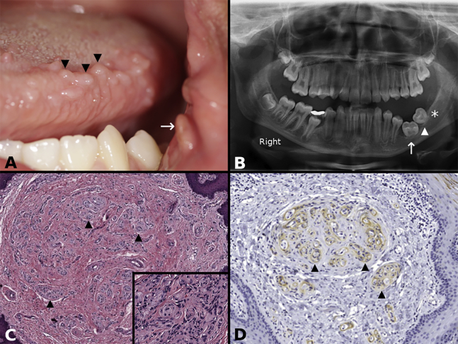
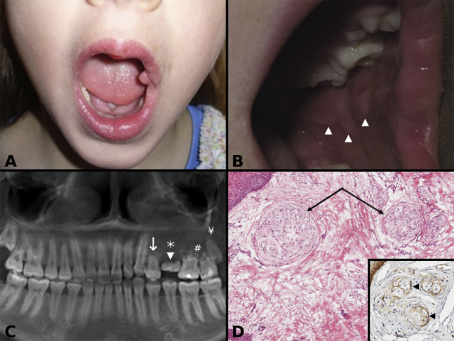
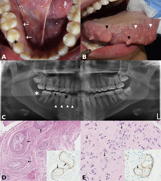
Individuals with orofacial asymmetry due to mucosal overgrowths, ipsilateral bone and dental aberrations with perineurial hyperplasia and/or perineuriomatous pseudo-onion bulb proliferations, comprise a recognizable clinical entity. In this article, we describe three individuals with this clinical entity and mosaic PIK3CA variants c.3140A>G (p. His1047Arg), c.328_330delGAA (p. Glu110del), and c.1353_1364del (p.Glu453_Leu456del). We conclude that the identification of these mosaic variants in individuals with orofacial asymmetry presenting histopathologically perineurial hyperplasia and/or intraneural pseudo-onion bulb perineurial cell proliferations supports the inclusion of this clinical entity in the PIK3CA-related overgrowth spectrum.

Keywords: PIK3CA; face; oral mucosa; overgrowth; perineurium; peripheral nerve; pseudo-onion bulbs.


Conflict of interest statement

The authors declare no competing interests.

Figures

Figure 1
Figure 1
Individual 1 (A) Clinical photograph of individual 1 showing multiple polypoid lesions on the left side of the tongue (three of many indicated with arrowheads) and a polyp at the oral commissure (arrow). (B) Orthopantomogram of individual 1 showing arrested root development of the permanent left first (arrow) and second (arrowhead) molars, with a missing third molar germ (asterisk). (C) Lower-power photomicrograph of individual 1 showing fibrous hyperplasia and multiple pseudo-onion bulb (perineurial cell) intraneural proliferations (three of many indicated with arrowheads) (hematoxylin and eosin stain). (Inset) High-power photomicrograph of individual 1 showing round structures representing pseudo-onion bulb (perineurial cell) intraneural proliferations (hematoxylin and eosin stain). (D) Epithelial membrane antigen (EMA) immunohistochemical stain decorates the perineurial cells of pseudo-onion bulb (perineurial cell) intraneural proliferations (three of many indicated with arrowheads; EMA with hematoxylin counterstain)
Figure 2
Figure 2
Individual 2 (A) Clinical photograph of individual 2 at age 3 years showing left side orofacial overgrowth, evident on the left side of the lip and tongue, and an oral commissural polyp. (B) Clinical photograph of individual 2 at age 10 years showing redundant tissue on the left buccal mucosa tissue (arrowheads). (C) Panoramic radiograph of individual 2 showing arrested development of the first left maxillary premolar (arrow), a retained primary second molar (arrowhead), an absent second premolar (asterisk), root resorption of the permanent molars (#), and a small third molar (¥). (D) Photomicrograph of individual 2 showing multiple pseudo-onion bulb (perineurial) intraneural proliferations (arrows) in a fibrous hyperplastic stroma (hematoxylin and eosin). (Inset) Epithelial membrane antigen (EMA) decorates (arrowheads) the perineurial cells of pseudo-onion bulb (perineurial) intraneural proliferations (EMA immunohistochemical stain with hematoxylin counterstain).
Figure 3
Figure 3
Individual 3 (A) Clinical photograph of individual 3 presenting with enlarged alveolus on the right side when compared to the left side (arrows). Black asterisk indicates missing right first premolar (see also C). (B) Clinical photograph of individual 3 showing polypoid lesions on the tongue (two of many; black arrowheads) and enlarged fungiform papillae (two of many; white arrowheads). (C) Panoramic radiograph of individual 3 showing absent right first mandibular premolar (white asterisk), small roots in the lower right teeth (white arrowheads), pulpal calcifications in the lower right premolar and molars (black arrowhead pointing to the pulpal calcification of the first permanent right molar), and missing third molar (black asterisk). The right body of the mandible is larger than the left (L), and there is apparent reduced bony trabeculation of the right side when compared to the left. (D) Photomicrograph of individual 3 showing perineurial hyperplasia (arrows) and peripheral nerve enlargement (hematoxylin and eosin). (Insert) Hyperplastic perineurium (arrows) decorated by epithelial membrane antigen (EMA; immunohistochemical stain with hematoxylin counterstain). (E) Photomicrograph of individual 3 showing multiple pseudo-onion bulb (perineurial) intraneural proliferations (arrows) (hematoxylin and eosin). (Inset) Pseudo-onion bulb (perineurial) intraneural proliferations decorated by epithelial membrane antigen (EMA; immunohistochemical stain with hematoxylin counterstain).

References

    1. Keppler-Noreuil K.M., Parker V.E., Darling T.N., Martinez-Agosto J.A. Somatic overgrowth disorders of the PI3K/AKT/mTOR pathway & therapeutic strategies. Am. J. Med. Genet. C. Semin. Med. Genet. 2016;172:402–421. - PMC - PubMed
    1. Rios J.J., Paria N., Burns D.K., Israel B.A., Cornelia R., Wise C.A., Ezaki M. Somatic gain-of-function mutations in PIK3CA in patients with macrodactyly. Hum. Mol. Genet. 2013;22:444–451. - PMC - PubMed
    1. Maclellan R.A., Luks V.L., Vivero M.P., Mulliken J.B., Zurakowski D., Padwa B.L., Warman M.L., Greene A.K., Kurek K.C. PIK3CA activating mutations in facial infiltrating lipomatosis. Plast. Reconstr. Surg. 2014;133:12e–19e. - PubMed
    1. Couto J.A., Konczyk D.J., Vivero M.P., Kozakewich H.P.W., Upton J., Fu X., Padwa B.L., Mulliken J.B., Warman M.L., Greene A.K. Somatic PIK3CA mutations are present in multiple tissues of facial infiltrating lipomatosis. Pediatr. Res. 2017;82:850–854. - PMC - PubMed
    1. Koutlas I.G., Scheithauer B.W. On pseudo-onion bulb intraneural proliferations of the non-major nerves of the oral mucosa. Head Neck Pathol. 2013;7:334–343. - PMC - PubMed

LinkOut - more resources