. 2022 Mar;9(9):e2105619.
doi: 10.1002/advs.202105619. Epub 2022 Jan 20.

Engineering a Rapid Insulin Release System Controlled By Oral Drug Administration

Affiliations

Engineering a Rapid Insulin Release System Controlled By Oral Drug Administration

Mohamed Mahameed et al. Adv Sci (Weinh). 2022 Mar.

Abstract

Rapid insulin release plays an essential role in maintaining blood-glucose homeostasis in mammalians. Patients diagnosed with type-I diabetes mellitus experience chronic and remarkably high blood-sugar levels, and require lifelong insulin injection therapy, so there is a need for more convenient and less invasive insulin delivery systems to increase patients' compliance and also to enhance their quality of life. Here, an endoplasmic-reticulum-localized split sec-tobacco etch virus protease (TEVp)-based rapamycin-actuated protein-induction device (RAPID) is engineered, which is composed of the rapamycin-inducible dimerization domains FK506 binding protein (FKBP) and FKBP-rapamycin binding protein fused with modified split sec-TEVp components. Insulin accumulation inside the endoplasmic reticulum (ER) is achieved through tagging its C-terminus with KDEL, an ER-retention signal, spaced by a TEVp cleavage site. In the presence of rapamycin, the split sec-TEVp-based RAPID components dimerize, regain their proteolytic activity, and remove the KDEL retention signal from insulin. This leads to rapid secretion of accumulated insulin from cells within few minutes. Using liver hydrodynamic transfection methodology, it is shown that RAPID quickly restores glucose homeostasis in type-1-diabetic (T1DM) mice treated with an oral dose of clinically licensed rapamycin. This rapid-release technology may become the foundation for other cell-based therapies requiring instantaneous biopharmaceutical availability.

Keywords: diabetes; endoplasmic reticulum; insulin; rapid release.


Conflict of interest statement

The authors declare no conflict of interest.

Figures
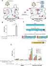
Figure 1
Figure 1
Design of fast‐release system using rapamycin as a signal input. A) Schematic illustration of RAPID in mammalian cells. The protein of interest (shown in blue) is C′‐terminally fused with a TEVp cleavage site (TCS) (shown in orange) and a KDEL retention signal (shown in green). In the absence of rapamycin, the system components are accumulated inside the ER through interaction with KDEL receptor (shown in red). Once rapamycin is added, RAPID components dimerize and regain catalytic functionality, cleaving the KDEL retention signal, and allowing the protein of interest to be secreted into the extracellular space. RAPID components are retained in the ER, as they contain an uncleavable KDEL signal that prevents their secretion. If KDEL‐bearing proteins escape the ER, they would be translocated back from the Golgi complex to the ER through COPI‐mediated retrieval vesicles (yellow). B) SEAP levels after 24 h in culture supernatants of HEK‐293 cells transfected with pMMH10 and pMMH51 plasmids, which encode SEAP‐TCS‐KDEL (PhCMV‐SEAP‐TCS‐KDEL‐pA) and intact sec‐TEVp‐KDEL (PhCMV‐sec‐TEVp‐KDEL‐pA) separately or in combination at a 1:1 ratio (50 ng each). Data are presented as means ± s.d. of n = 6 biologically independent samples. *** p < 0.001. C) Schematic illustration of the protein components of the RAPID expression system. Sec‐TEVp was split into two parts; sec‐TEVp‐N′1–118 and sec‐TEVp‐C′119–245, which are fused with FKBP and FRB, respectively. Both constructs are tagged with a C′‐terminal KDEL retention signal and an N′‐terminal calreticulin secretion signal (SS), which directs them to the ER. To enhance the dimerization efficiency, a flexible linker (GGGGS)2 is inserted between FKBP and sec‐TEVp‐N′1–118. D) SEAP levels in culture supernatants of HEK‐293 cells transfected with pMMH26 and PMMH27 plasmids, which encode SS‐FKBP‐(GGGGS)2‐ssTEVp1–118‐KDEL (PhCMV‐SS‐FKBP‐(GGGGS)2‐ssTEVp1–118‐KDEL‐pA) and SS‐FRB‐ssTEVp119–245‐KDEL (PhCMV‐SS‐FRB‐ssTEVp119–245‐KDEL‐pA) (5 ng each), and different amounts of pMMH10 plasmid, which encodes SEAP‐TSC‐KDEL (PhCMV‐SEAP‐TCS‐KDEL‐pA). At 24 h after transfection, the medium was replaced with 30 µL of fresh medium containing either DMSO or rapamycin (100 nm) and 20 µL aliquots were collected for analysis at different time points. Data are presented as means ± s.d. of n = 3 biologically independent samples. *** p < 0.001.
Figure 2
Figure 2
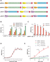
Engineering an efficient RAPID‐based expression system in a single tri‐cistronic expression vector. A) To find the optimal ratios among different parts of RAPID expression system, systematic engineering of different plasmids using PhEF1a, PEF1a core, and PhPGK was performed. B) SEAP levels in the supernatant of HEK‐293 cells transfected with the different constructs presented previously (100 ng of each plasmid separately). At 24 h after transfection, the medium was replaced with 30 µL of fresh medium containing either DMSO or rapamycin (100 nm) and 20 µL aliquots were collected for analysis at different time points. Data are presented as means ± s.d. of n = 3 biologically independent samples. *** p < 0.001. C) SEAP level in the supernatant of HEK‐293 cells transfected with different amounts (5–100 ng) of pBS971. 24 h after transfection, the culturing medium was replaced with 30 µL of 100 nm rapamycin‐containing medium (or with DMSO as negative control) and SEAP expression was profiled for after 50 min. Data are presented as means ± s.d. of n = 6 biologically independent samples. D) Dose‐response analysis using pBS97 plasmid, which was found to be the most efficient plasmid with minimal leakiness. HEK‐293 cells were transfected with 5 ng of pBS97. At 24 h after transfection, the medium was replaced with 30 µL of fresh medium containing different concentrations of rapamycin and 20 µL aliquots were collected for analysis. Data are presented as means ± s.d. of n = 3 biologically independent samples. E) Comparison of RAPID and TET‐ON expression systems. Cells were transfected either with pBS971 or with plasmids encoding PhCMV*‐1‐SEAP (PhCMV*‐1‐SEAP‐pA, pTS1017) and M2rtTA (PUbc‐M2rtTA‐pA). At 20 h after transfection, the culture medium was replaced with fresh medium and the cells were left for 24 h to recover and to accumulate the system components. The medium was replaced with 30 µL of fresh medium containing either doxycycline (2 µg mL−1) or rapamycin (100 nm) and 20 µL aliquots were collected for analysis at different time points. Data are presented as means ± s.d. of n = 6 biologically independent samples. *** p < 0.001.
Figure 3
Figure 3
Evaluation of RAPID performance in mice in vivo. A) Top: a schematic illustration of the genetic components of pMMH122, which encodes RAPID‐mediated expression of NanoLuc luciferase (NLuc). Bottom: values of relative luminescence units (RLU) in culture supernatants of HEK‐293 cells transfected with different amounts of pMMH122. At 24 h after transfection, the medium was replaced with 30 µL of fresh medium containing either DMSO or rapamycin (100 nm) and 7.5 µL aliquots were collected for analysis after 50 min. Data are presented as means ± s.d. of n = 4 biologically independent samples. B) RLU in the plasma of healthy WT mice following rapamycin treatment at different doses. At 24 h following hydrodynamic transfection of pMMH122, mice were given with different amounts of rapamycin by oral gavage. Blood samples were collected at different time points and RLU values were quantified. Data are presented as means ± s.d. of n = 5. C) RLU in the plasma of WT mice following multiple rapamycin treatments. At 24 h following hydrodynamic transfection of pMMH122, mice were treated three times with oral rapamycin (0.2 mg kg−1 each time) at 12 h intervals for a period of 36 h. Each point represents the mean of plasma RLU values before or 1 h after rapamycin treatment. Data are presented as means ± s.d. of n = 5.
Figure 4
Figure 4
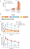
Fast insulin release in T1DM in response to oral administration of rapamycin. A) SEAP levels in insulin‐activity bioassay as a readout of insulin activity. To produce fast‐release insulin, HEK‐293 cells were transfected either with INS‐TCS‐KDEL (pMMH50) in combination with intact sec‐TEVp (pMMH51), or with pcDNA3.1 (+) as a negative control. At 12 h after transfection, the culture medium was replaced with fresh DMEM and incubation was continued for 24 h. The supernatant was used for insulin‐activity bioassay, and SEAP measurements were performed after 24 h (see insulin quantification in the Experimental Section). Clinical insulin (1 µg L−1) was used as a positive control. Data are presented as means ± s.d. of n = 4 biologically independent samples. *** p < 0.001. B) The structures of tetra‐cistronic insulin RAPID‐based expression plasmids used for in vivo hydrodynamic transfection of the liver (pMMH68). C) Mealtime challenge in T1DM or healthy WT mice following rapamycin treatment. At 24 h following hydrodynamic transfection of pMMH68, mice were fasted for 8 h and treated with oral rapamycin gavage (0.2 mg kg−1) or vehicle plus to intraperitoneal glucose injection (125 g kg−1). Blood glucose levels were then measured at different time points. Normal glycemia values are indicated by an orange band. Data are presented as means ± s.d. of n = 5. *** p < 0.001. D) Blood insulin profiles in mice from the previous experiment (Figure 3C) using cell culture‐based bioassay. Data are presented as means ± s.d. of n = 5. *** p < 0.001.

References

    1. Petersen M. C., Shulman G. I., Physiol. Rev. 2018, 98, 2133. - PMC - PubMed
    1. Fargion S., Dongiovanni P., Guzzo A., Colombo S., Valenti L., Fracanzani A. L., Aliment. Pharmacol. Ther. 2005, 22, 61. - PubMed
    1. Seino S., Shibasaki T., Minami K., J. Clin. Invest. 2011, 121, 2118. - PMC - PubMed
    1. Komatsu M., Takei M., Ishii H., Sato Y., J. Diabetes Investig. 2013, 4, 511. - PMC - PubMed
    1. DiMeglio L. A., Evans‐Molina C., Oram R. A., Lancet 2018, 391, 2449. - PMC - PubMed

Publication types

LinkOut - more resources