. 2022 Feb 22;50(3):1517-1530.
doi: 10.1093/nar/gkac011.

TRIM21 suppresses CHK1 activation by preferentially targeting CLASPIN for K63-linked ubiquitination

Affiliations

TRIM21 suppresses CHK1 activation by preferentially targeting CLASPIN for K63-linked ubiquitination

Xuefei Zhu et al. Nucleic Acids Res. .

Abstract

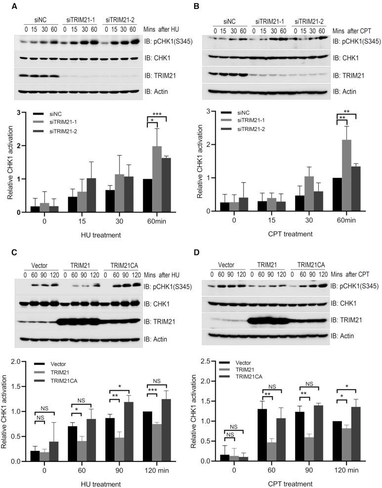
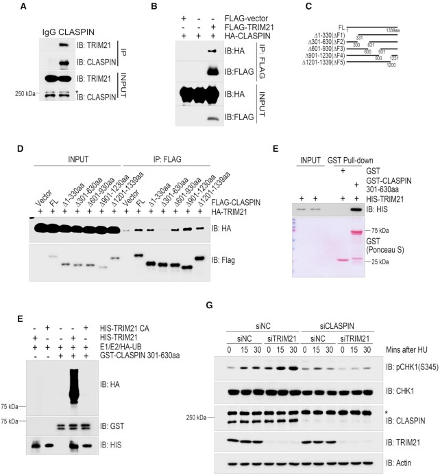
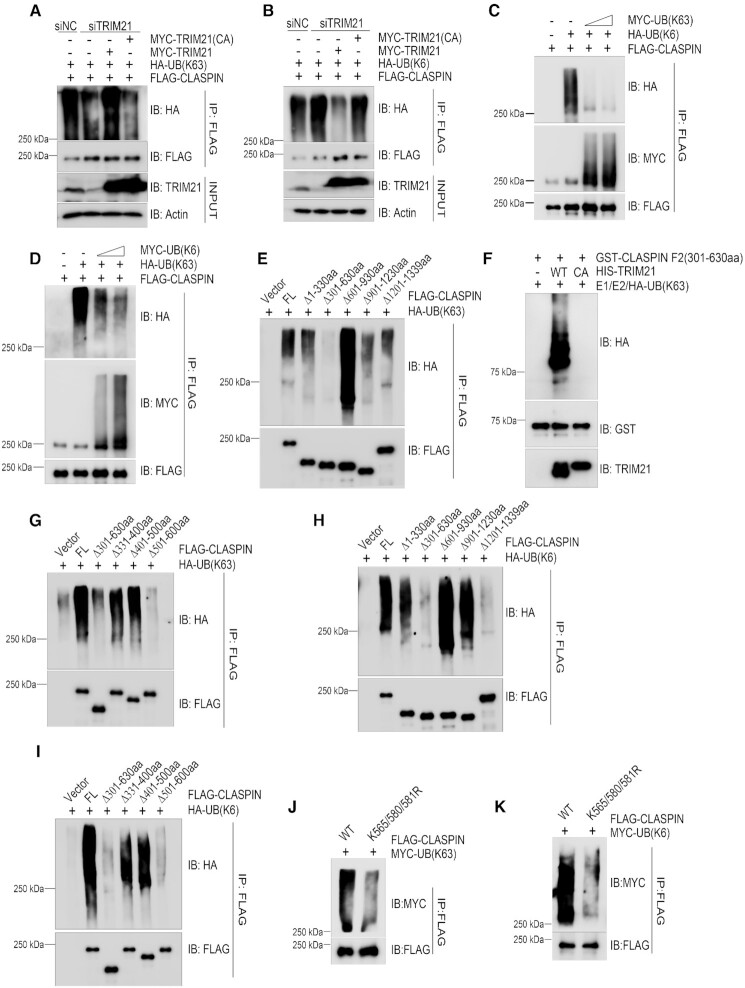
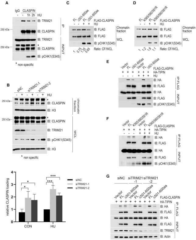
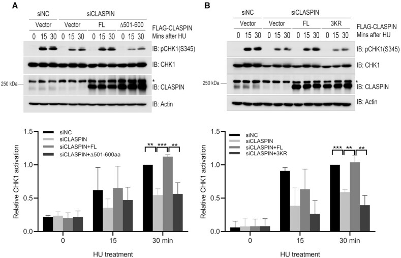
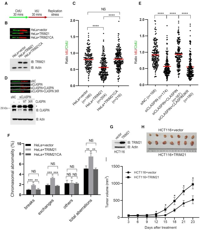
Expression of the E3 ligase TRIM21 is increased in a broad spectrum of cancers; however, the functionally relevant molecular pathway targeted by TRIM21 overexpression remains largely unknown. Here, we show that TRIM21 directly interacts with and ubiquitinates CLASPIN, a mediator for ATR-dependent CHK1 activation. TRIM21-mediated K63-linked ubiquitination of CLASPIN counteracts the K6-linked ubiquitination of CLASPIN which is essential for its interaction with TIPIN and subsequent chromatin loading. We further show that overexpression of TRIM21, but not a TRIM21 catalytically inactive mutant, compromises CHK1 activation, leading to replication fork instability and tumorigenesis. Our findings demonstrate that TRIM21 suppresses CHK1 activation by preferentially targeting CLASPIN for K63-linked ubiquitination, providing a potential target for cancer therapy.


Figures

Figure 1.
Figure 1.
TRIM21 negatively regulated CHK1 activation upon replication stress. (A) HeLa cells were transfected with a negative control siRNA (siNC) or siRNA targeting 3’UTR of TRIM21 (siTRIM21-1 and siTRIM21-2) for 48 h. Transfectants were treated with 2 mM HU for the indicated time and total cell lysates were harvested for immunoblotting with antibodies as indicated. Statistical analysis (t-test) of signal intensity from three independent experiments was shown in the lower panel. *P < 0.05; ***P < 0.001. (B) HeLa cells were treated as described in (A) except that HU treatment was replaced with CPT (1 μM) treatment. **P < 0.01. (C) HeLa cells overexpressing TRIM21 or catalytically inactive mutant TRIM21CA were treated with 2 mM HU for the indicated time before total cell lysates were extracted for immunoblotting with antibodies as indicated. Statistical analysis (t-test) of signal intensity from three independent experiments was shown in the lower panel. *P < 0.05; **P < 0.01; ***P < 0.001. (D) HeLa cells overexpressing TRIM21 or TRIM21CA were treated with 1 μM CPT for the indicated time before total cell lysates were extracted for immunoblotting with antibodies as indicated. *P < 0.05; **P < 0.01.
Figure 2.
Figure 2.
TRIM21 ubiquitinated CLASPIN and regulated CLASPIN dependent CHK1 activation. (A) HEK293T cells were lysed and incubated with protein A agarose conjugated with normal rabbit IgG or anti-CLASPIN antibody for immunoprecipitation, followed by immunoblotting with the indicated antibodies. *, non-specific signal. (B) HEK293T cells co-transfected with HA-CLASPIN and FLAG-TRIM21 were lysed and incubated with anti-FLAG M2 agarose, the immunoprecipitates were then examined by immunoblotting with antibodies against HA and FLAG. (C) Schematic of the CLASPIN deletion mutants: F1:1–330aa, F2:301–630aa, F3:601–930aa, F4:901–1230aa, F5:1201–1339aa. (D) Whole cell lysates extracted from HEK293T cells co-transfected with HA-TRIM21 and wildtype or mutant FLAG-CLASPIN were incubated with anti-FLAG M2 agarose for immunoprecipitation, followed by immunoblotting with the indicated antibodies. (E) Bacterially-purified HIS-TRIM21 and GST-CLASPIN301-630aa/GST peptides immobilized on Glutathione Sepharose 4B beads were mixed and incubated for 4 h. The HIS-TRIM21 complex was examined by immunoblotting. GST-CLASPIN301-630aa/GST were directly visualized by Ponceau S staining. (F) In vitro ubiquitination assays were performed by incubating HIS-TRIM21 or its E3 ligase inactive mutant and GST-CLASPIN301-630aa in the presence of E1, E2 and HA-ubiquitin at 30°C for 1 h, followed by GST-pulldown and analysis by immunoblotting with the indicated antibodies. (G) HeLa cells were transfected with a negative control siRNA or an siRNA targeting TRIM21 or CLASPIN as indicated for 48 hours. Then, whole cell lysates were collected after being treated with 2 mM HU or a mock treatment for the indicated times. CHK1 activation was examined by immunoblotting with an antibody specifically recognizing phosphorylated CHK1 at Ser345. The expression levels of CLASPIN, TRIM21 and CHK1 were also examined. *Non-specific signal. Actin: loading control.
Figure 3.
Figure 3.
TRIM21 ubiquitinated CLASPIN with K63 linkage that counteracted its K6-linkage ubiquitination. (A, B) HEK293T cells were transfected with a negative control or a TRIM21-specific siRNA for 24 h. Then, the cells were co-transfected with FLAG-CLASPIN and HA-ubiquitin K63-only (as in A) or HA-ubiquitin K6-only (as in B) together with MYC-TRIM21 or MYC-TRIM21 E3 ligase inactive mutant as indicated. After 24 h, all the cell lines were lysed and subjected to de-naturing immunoprecipitation using anti-FLAG M2 agarose. The immunoprecipitates were examined by immunoblotting with the indicated antibodies. (C, D) HEK293T cells were transfected with FLAG-CLASPIN, and HA-ubiquitin K6-only along with increasing doses of MYC-ubiquitin K63-only expressing plasmids (as in C) or HA-ubiquitin K63-only along with increasing doses of MYC-ubiquitin K6-only expressing plasmids (as in D). After 24 h, the cells were lysed and analyzed by de-naturing immunoprecipitation using anti-FLAG M2 agarose, followed by immunoblotting with the indicated antibodies. (E) HEK293T cells were co-transfected with HA-ubiquitin K63-only and full-length FLAG-CLASPIN or its deletion mutants, and then analyzed by de-naturing immunoprecipitation and immunoblotting with the indicated antibodies. (F) In vitro ubiquitination assays were performed by incubating HIS-TRIM21 or TRIM21CA with GST-CLASPIN 301–630aa in the presence of E1, E2 and HA-ubiquitin-K63 only (HA-UB(K63)) at 30°C for 1 h, followed by GST-pulldown and analysis by immunoblotting with the indicated antibodies. (G) K63-linked ubiquitination of wildtype CLASPIN and its deletion mutants was examined as described in (E). (H) HEK293T cells were co-transfected with HA-ubiquitin K6-only and full-length FLAG-CLASPIN or its deletion mutants, and analyzed by denaturing immunoprecipitation and immunoblotting with the indicated antibodies. (I) HEK293T cells were transfected with the indicated plasmids and analyzed by de-naturing immunoprecipitation and immunoblotting with the indicated antibodies. (J, K) HEK293T cells were co-transfected with MYC-ubiquitin K63-only (as in J) or MYC-ubiquitin K6-only (as in K) and wildtype FLAG-CLASPIN or its K565/580/581R mutant. The samples were analyzed by de-naturing immunoprecipitation and immunoblotting with the indicated antibodies.
Figure 4.
Figure 4.
TRIM21 negatively regulated the chromatin loading of CLASPIN and its interaction with TIPIN. (A) HEK293T cells were treated with 2 mM HU for the indicated times, total cell lysates were extracted and subjected to immunoprecipitation and immunoblotting with antibodies as indicated. *, non-specific signal. (B) HeLa cells were transfected with siNC or siTRIM21 for 24 h and subjected to double thymidine blocks to synchronize cells in G1/S transition followed by a release for 2 h. Transfectants were untreated or treated with 2 mM HU for 1 h before chromatin fractionation was performed. The chromatin-enriched fraction was analyzed by immunoblotting with the indicated antibodies. * Non-specific signal. Statistical analysis (t-test) of signal intensity from three independent experiments was shown in the lower panel. *P < 0.05; ***P < 0.001. (C) HEK293T cells were transfected with FLAG-CLASPIN or the FLAG-CLASPINΔ501–600 mutant. After 24 h, the cells were treated with 2 mM HU or a mock treatment for 1 h and subjected to chromatin-bound protein extraction. The protein levels of FLAG-CLASPIN or FLAG-CLASPINΔ501–600aa in the chromatin fraction and whole cell lysate were detected by immunoblotting, and relative chromatin loading was analyzed by comparing the ratio between the chromatin fraction and the whole cell lysate. (D) HEK293T cells were transfected with FLAG-CLASPIN or the FLAG-CLASPIN K565/580/581R. Then cells were treated and analyzed by immunoblotting as described in (C). (E) HEK293T cells were transfected with HA-TIPIN and FLAG-CLASPIN or its Δ501–600aa mutant. After 24 h, the cells were treated with 2 mM HU or a mock treatment for 1 h, and subjected to immunoprecipitation using anti-FLAG M2 agarose, followed by immunoblotting with the indicated antibodies. (F) HEK293T cells were transfected with HA-TIPIN and FLAG-CLASPIN or its K565/580/581R mutant. After 24 h, the cells were treated with 2 mM HU or a mock treatment for 1 h and subjected to immunoprecipitation using anti-FLAG M2 agarose, followed by immunoblotting with the indicated antibodies. (G) HEK293T cells were transfected with a negative control or different TRIM21 specific siRNAs for 24 h. Then the cells were transfected with HA-TIPIN and FLAG-CLASPIN or its Δ501–600aa mutant, and analyzed by immunoprecipitation using anti-FLAG M2 agarose followed by immunoblotting with the indicated antibodies.
Figure 5.
Figure 5.
TRIM21-mediated CLASPIN ubiquitination suppressed CHK1 activation. (A) HEK293T cells were transfected with a negative control or a siRNA targeting for the 3’UTR of CLASPIN (siCLASPIN) for 24 h. Then, the cells were transfected with FLAG-CLASPIN or FLAG-CLASPINΔ501–600aa as indicated and incubated for a further 24 h. Then the cells were treated with 2 mM HU or a mock treatment before the whole cell lysates were collected and analyzed by immunoblotting. Activation of CHK1 was examined by immunoblotting with an antibody specifically recognizing phosphorylated CHK1 at Ser345. The CHK1 and CLASPIN expression levels were also examined. Actin: loading control. *, non-specific signal. Statistical analysis (t-test) of signal intensity from three independent experiments was shown in the lower panel. **P < 0.01; ***P < 0.001. (B) HEK293T cells were transfected with a negative control or a CLASPIN-specific siRNA for 24 h. Then, the cells were transfected with FLAG-CLASPIN or FLAG-CLASPIN K565/580/581R (3KR) mutant as indicated, and incubated for a further 24 h. Then the cells were treated and examined as in (A). *, non-specific signal. Statistical analysis (t-test) of signal intensity from three independent experiments was shown in the lower panel. **P < 0.01; ***P < 0.001.
Figure 6.
Figure 6.
Overexpression of TRIM21 led to genome instability and tumorigenesis. (A) Schematic of the DNA fiber assay examining stalled replication fork stability. Cells were sequentially labeled with 40 μM CldU and 100 μM IdU for 30 min each, followed by treatment with 5 mM hydroxyurea and 5 μM aphidicolin to block replication fork progression. (B, C) HeLa cells or HeLa cells overexpressing either TRIM21 or TRIM21(CA) were treated as described in (A); the DNA fibers were spread and subjected to immunofluorescence using anti-BrdU antibodies specifically recognizing CldU or IdU. (B) Representative images of the DNA fibers and the TRIM21 expression level. (C) For each group, a IdU/CldU ratio of > 150 individual DNA fibers is presented and the mean IdU/CldU ratio (marked as the red line) is shown. ****, t-test, P < 0.0001. (D, E) HeLa cells and HeLa cells expressing exogenous CLASPIN or ubiquitination-defective mutant CLASPIN(K565/580/581R) were transfected with siNC or siCLASPIN for 48 h before subjected to DNA fiber assay as described in (A) and immunoblotting with antibodies as indicated. Representative images of the DNA fibers and the CLASPIN expression levels are shown in (D). * Non-specific signal. (E) For each group, a IdU/CldU ratio of >150 individual DNA fibers is presented and the mean IdU/CldU ratio (marked as the red line) are shown in (E). ****, t-test, P < 0.0001. (F) HeLa cells stably overexpressing TRIM21 or its E3 ligase inactive mutant were subjected to metaphase spread assay; >1500 metaphase chromosomes were analyzed for every cell line. The error bars represent the SD, n = 3. t-test, **P < 0.01; ***P < 0.001. (G–I) 6 × 106 HCT116 cells overexpressing empty vector or TRIM21 were implanted into nude mice, and the tumor volume was monitored at the indicated times. The TRIM21 expression level was examined (G). Tumor images (H) and quantification results are shown (I). n = 7, mean tumor volume ± SEM, t-test, *P < 0.05.

Similar articles

Cited by

References

    1. Hoeijmakers J.H. DNA damage, aging, and cancer. N. Engl. J. Med. 2009; 361:1475–1485. - PubMed
    1. Zeman M.K., Cimprich K.A.. Causes and consequences of replication stress. Nat. Cell Biol. 2014; 16:2–9. - PMC - PubMed
    1. Ghosal G., Chen J.. DNA damage tolerance: a double-edged sword guarding the genome. Transl. Cancer Res. 2013; 2:107–129. - PMC - PubMed
    1. Jones R.M., Petermann E.. Replication fork dynamics and the DNA damage response. Biochem. J. 2012; 443:13–26. - PubMed
    1. Saldivar J.C., Cortez D., Cimprich K.A.. The essential kinase ATR: ensuring faithful duplication of a challenging genome. Nat. Rev. Mol. Cell Biol. 2017; 18:622–636. - PMC - PubMed

Publication types

MeSH terms