. 2021 Dec 26;29(1):94-110.
doi: 10.3390/curroncol29010008.

The Neurokinin-1 Receptor Is a Target in Pediatric Rhabdoid Tumors

Affiliations

The Neurokinin-1 Receptor Is a Target in Pediatric Rhabdoid Tumors

Julian Kolorz et al. Curr Oncol. .

Abstract

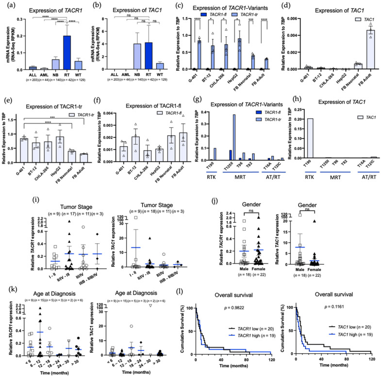
Rhabdoid tumors (RT) are among the most aggressive tumors in early childhood. Overall survival remains poor, and treatment only effectively occurs at the cost of high toxicity and late adverse effects. It has been reported that the neurokinin-1 receptor/ substance P complex plays an important role in cancer and proved to be a promising target. However, its role in RT has not yet been described. This study aims to determine whether the neurokinin-1 receptor is expressed in RT and whether neurokinin-1 receptor (NK1R) antagonists can serve as a novel therapeutic approach in treating RTs. By in silico analysis using the cBio Cancer Genomics Portal we found that RTs highly express neurokinin-1 receptor. We confirmed these results by RT-PCR in both tumor cell lines and in human tissue samples of various affected organs. We demonstrated a growth inhibitory and apoptotic effect of aprepitant in viability assays and flow cytometry. Furthermore, this effect proved to remain when used in combination with the cytostatic cisplatin. Western blot analysis showed an upregulation of apoptotic signaling pathways in rhabdoid tumors when treated with aprepitant. Overall, our findings suggest that NK1R may be a promising target for the treatment of RT in combination with other anti-cancer therapies and can be targeted with the NK1R antagonist aprepitant.

Keywords: NK-1 receptor; NK-1 receptor antagonist; apoptosis; cancer; rhabdoid tumor; substance P.


Conflict of interest statement

S.K. has received honoraria from Novartis, TCR2 and GSK. S.K. is an inventor of several patents in the field of immuno-oncology. S.K. received research support from TCR2 Inc and Arcus Bioscience for work unrelated to this manuscript.

Figures

Figure 1
Figure 1
Expression of TACR1 and TAC1 and biological and clinical parameters of rhabdoid tumors. (a,b) mRNA expression of TACR1 and TAC1 in ALL (n = 203), AML (n = 44), NB (n = 140), RT (n = 42), and WT (n = 129). Relative expression was correlated to the (i) tumor stage by the Neoplasm American Joint Committee on Cancer (I-II n = 9; II/IV-III n = 17 for TACR1, n = 18 for TAC1; III/IV n = 11; IIIB-IIIB/IV n = 3), (j) gender (Male n = 18; Female n = 22), (k) age at diagnosis (months) (< 6 months n = 9; 6–12 months n = 15; 12–18 months n = 5; 18–24 months n = 3; 24–40 months n = 2; > 30 months n = 6), (l) overall survival (months) (TACR1 low n = 20; TACR1 high n = 19; TAC1 low (n = 20), TAC1 high n = 19), and (cf) mRNA expression of TACR1-fl, TACR1-tr, and TAC1 in G-401, BT-12, CHLA-266, HepG2, and in FB Neonatal (=fibroblasts neonatal, PCS-201-120) and in FB Adult (=fibroblasts adult, PCS-201-012) normalized to the housekeeping gene TBP. (g,h) mRNA expression of TACR1-fl, TACR1-tr and TAC1 in tumor samples normalized to TBP. Results are expressed as the mean ± standard error of the mean (SEM). All statistical comparisons were made with an unpaired parametric t-test comparing two groups, an ordinary one-way ANOVA or a Tukey’s multiple comparison test, with a single pooled variance. ns = not significant. p < 0.05 (*), p < 0.001 (***) and p < 0.0001 (****) for all comparisons.
Figure 2
Figure 2
Cell proliferation analysis of G-401, BT-12, CHLA-266, HepG2, FB Neonatal, and FB Adult upon treatment with different compounds. (a) MTT assay measuring cell viability upon treatment with aprepitant (10–100 μM) for 48 h. (b) MTT assay measuring cell viability upon treatment with cisplatin (0.02–100 μM) for 48 h. (c) Combination MTT assay measuring cell viability upon treatment of aprepitant (aprepitant concentrations for G-401 and BT-12, 50 µM; CHLA-266, 40 µM; HepG2 15 µM), cisplatin (Cis, 20 μM) and aprepitant + cisplatin (Combi) for 48 h. (d) MTT assay measuring cell viability upon treatment of aprepitant (G-401 and BT-12, 50 µM; CHLA-266, 40 µM; HepG2 15 µM) and stimulation with Substance P (SP, 200 nM) and combination of Apre and SP. Pooled data of three independent experiments. Results are expressed as the mean ± standard error of the mean (SEM). All statistical comparisons were made with an ordinary one-way ANOVA or a Tukey’s multiple comparison test, with a single pooled variance. p < 0.05 (*), p < 0.01 (**), p < 0.001 (***) and p < 0.0001 (****) for all comparisons.
Figure 3
Figure 3
(a) Determination of apoptotic cell populations of G-401, BT-12, CHLA-266, and HepG2 was performed by staining with annexin V and fixable viability dye (FVD) and assessed through fluorescence-activated cell sorting. Cells were treated with aprepitant (aprepitant concentrations for G-401 and BT-12, 50 µM; CHLA-266, 40 µM; HepG2, 15 µM), cisplatin (Cis, 20 μM) and aprepitant + cisplatin (Combi) for 48 h. DMSO was used as treatment control. Shown is one representative of three experiments. Numbers represent the percentages of the cell populations; black: viable; blue: apoptotic. (b) Quantification of viable and apoptotic cell populations. Shown are results from three independent experiments (n = 3).
Figure 4
Figure 4
(a) Western blot analysis of G-401, BT-12 and CHLA-266 upon treatment with aprepitant (G-401 and BT-12, 50 µM; CHLA-266, 40 µM), cisplatin (CIS 20 µM) and aprepitant + cisplatin (COMBI) (48 h). DMSO used as a treatment control. Blots are representative of n = 2. (b) Densitometry analysis of Western blots. Relative protein expression levels were normalized to alpha tubulin expression.

References

    1. Geller J.I., Roth J.J., Biegel J.A. Biology and Treatment of Rhabdoid Tumor. Crit. Rev. Oncog. 2015;20:199–216. doi: 10.1615/CritRevOncog.2015013566. - DOI - PMC - PubMed
    1. Chi S.N., Zimmerman M.A., Yao X., Cohen K.J., Burger P., Biegel J.A., Rorke-Adams L.B., Fisher M.J., Janss A., Mazewski C., et al. Intensive multimodality treatment for children with newly diagnosed CNS atypical teratoid rhabdoid tumor. J. Clin. Oncol. 2009;27:385–389. doi: 10.1200/JCO.2008.18.7724. - DOI - PMC - PubMed
    1. Fazlollahi L., Hsiao S.J., Kochhar M., Mansukhani M.M., Yamashiro D.J., Remotti H.E. Malignant Rhabdoid Tumor, an Aggressive Tumor Often Misclassified as Small Cell Variant of Hepatoblastoma. Cancers. 2019;11:1992. doi: 10.3390/cancers11121992. - DOI - PMC - PubMed
    1. Chao M.F., Su Y.F., Jaw T.S., Chiou S.S., Lin C.H. Atypical teratoid/rhabdoid tumor of lumbar spine in a toddler child. Spinal Cord Ser. Cases. 2017;3:16026. doi: 10.1038/scsandc.2016.26. - DOI - PMC - PubMed
    1. Bourdeaut F., Freneaux P., Thuille B., Bergeron C., Laurence V., Brugieres L., Verite C., Michon J., Delattre O., Orbach D. Extra-renal non-cerebral rhabdoid tumours. Pediatr. Blood Cancer. 2008;51:363–368. doi: 10.1002/pbc.21632. - DOI - PubMed

Publication types

MeSH terms

LinkOut - more resources