Review
. 2022 Jan 8;12(2):145.
doi: 10.3390/ani12020145.

Integrity of the Intestinal Barrier: The Involvement of Epithelial Cells and Microbiota-A Mutual Relationship

Affiliations
Review

Integrity of the Intestinal Barrier: The Involvement of Epithelial Cells and Microbiota-A Mutual Relationship

Małgorzata Gieryńska et al. Animals (Basel). .

Abstract

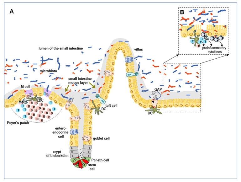
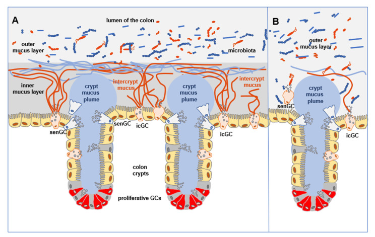
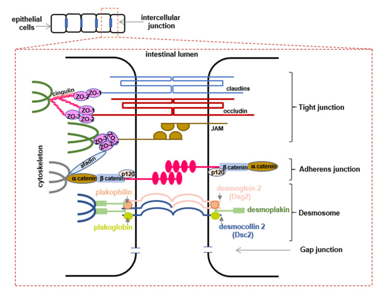
The gastrointestinal tract, which is constantly exposed to a multitude of stimuli, is considered responsible for maintaining the homeostasis of the host. It is inhabited by billions of microorganisms, the gut microbiota, which form a mutualistic relationship with the host. Although the microbiota is generally recognized as beneficial, at the same time, together with pathogens, they are a permanent threat to the host. Various populations of epithelial cells provide the first line of chemical and physical defense against external factors acting as the interface between luminal microorganisms and immunocompetent cells in lamina propria. In this review, we focus on some essential, innate mechanisms protecting mucosal integrity, thus responsible for maintaining intestine homeostasis. The characteristics of decisive cell populations involved in maintaining the barrier arrangement, based on mucus secretion, formation of intercellular junctions as well as production of antimicrobial peptides, responsible for shaping the gut microbiota, are presented. We emphasize the importance of cross-talk between gut microbiota and epithelial cells as a factor vital for the maintenance of the homeostasis of the GI tract. Finally, we discuss how the imbalance of these regulations leads to the compromised barrier integrity and dysbiosis considered to contribute to inflammatory disorders and metabolic diseases.

Keywords: Paneth cells; epithelial cells; gastrointestinal tract; goblet cells; microbiota.


Conflict of interest statement

The authors declare no conflict of interest.

Figures

Figure 1
Figure 1
A representative schematic of villi, crypts of Lieberkühn and Peyer’s patches of the small intestine (according to [9]). (A) Constantly dividing stem cells, located at the base of the crypts of Lieberkühn, intercalating with Paneth cells, give rise to the progenitor cells differentiating into mature intestinal epithelial cells. Paneth cells, due to the proximity of stem cells and secretion of specific mediators, are considered to be protectors of this population. Moreover, they are the major source of antimicrobial peptides found in the mucus of the small intestine, and are responsible for controlling the gut microbiota, preventing its overgrowth, and inhibiting inflammation. Enterocytes, absorptive cells, comprise the most numerous populations, and together with goblet cells, are found in the crypt-villus axis, and follicle-associated epithelium (FAE). Goblet cells are responsible for the formation of the mucus layer covering the small intestine epithelium, due to the secretion of mucin MUC2. The mucus barrier in the small intestine is penetrable for microbiota. Goblet cells also facilitate luminal antigen sampling and their transport to the lamina propria located dendritic cells via goblet cell-associated passages (GAPs). FAE overlying Peyer’s patch, contain scattered, devoid of mucus layer M cells, which participate in direct sampling of luminal antigens, translocating them to the basolateral site where antigen presenting cells, located in lamina propria, can capture, process, and present them, inducing tolerance or antigen-specific immune response. Tuft cells and enteroendocrine cells are sparse in the intestinal lining. They are involved in protection against helminth infection or secrete peptides and hormones which stimulate, among others, intestinal peristalsis. (B) Disturbance of the mucus barrier results in penetration and direct contact with epithelial cells with microorganisms. This results in inflammatory response induction, recruitment of neutrophils, induction of dendritic cells and macrophages localized in lamina propria, stimulation of T cells and B cells, and secretion of proinflammatory cytokines. Inflammatory response leads to the injury of epithelial cells.
Figure 2
Figure 2
A representative schematic of colon crypts (according to [32]). (A) Colonocytes are the most numerous epithelial cell population of the colon. Constantly dividing stem cells differentiate into proliferative goblet cells that give rise to diverse populations of goblet cells that participate in the formation of mucus barrier, due to secretion of MUC2. The colon mucus barrier is formed of the inner mucus layer, free of microbiota and outer mucus layer, penetrable for microorganisms. Thick crypt mucus plumes, impermeable for bacteria, shield stem cells area, are secreted by crypt goblet cells among which, at the entrance to the crypt are located sentinel goblet cells (senGCs). Upon TLR ligand stimulation, senGCs form NLRP6 inflammasome, and the production of reactive oxygen species occurs. This will lead to the release of Ca2+ that passed through gap junctions to neighboring cells, inducing mucus release. Activated senGCs are expelled to the lumen. Between crypts lie highly differentiated intercrypt goblet cells (inGCs), secreting intercrypt mucus, penetrable for small molecules. Intercrypt mucus and crypt mucus plume form the colon inner mucus layer. Together, populations of goblet cells form a network that protects epithelium against microbial colonization. Any disruption of inGCs results in epithelial exposure to bacteria and the possible development of colitis. (B) The diminished number of inGCs due to the increased cell shedding to the lumen results in altering the mucus barrier in the colon. The only outer mucus layer is present, which is penetrable for microorganisms, which can contribute to colitis development.
Figure 3
Figure 3
A representative schematic of the intercellular junctions in the intestinal epithelium (according to [5,54,55]). The intestinal epithelium forms a barrier maintained by a structure composed of three junctions: tight junctions (TJs), adherens junctions (AJs), and desmosomes. TJs are generated by membrane proteins, claudins, occludin, and junctional adhesion molecules (JAM) that interact in the paracellular space with proteins on adjacent cells, completely closing the apical intercellular space, blocking microbial penetration, and regulating the selective passage of macromolecules. TJ complex is connected to the actin cytoskeleton through adaptor proteins zonula occludens-1 (ZO-1), ZO-2, ZO-3, as well as other proteins (cingulin, afadine). The lower junctional complexes, AJs, and desmosomes provide adhesive and mechanical properties that contribute to the barrier functions. AJs are created mainly by membrane E-cadherins that form homophilic cell-cell interactions and are intracellularly connected with catenins (α-catenin, β-catenin, p120 catenin) through which they link E-cadherins with a cytoskeleton. Desmosomes are composed of desmoglein 2 and desmocollin 2, forming homo- and heterophilic interactions. They are linked to plakophilins and plakoglobin via the cytoplasmatic domain. These in turn are connected to the intermediate filaments by desmoplakin.

References

    1. Kim S.-H., Lee K.-Y., Jang Y.-S. Mucosal Immune System and M Cell-targeting Strategies for Oral Mucosal Vaccination. Immune Netw. 2012;12:165. doi: 10.4110/in.2012.12.5.165. - DOI - PMC - PubMed
    1. Sansonetti P.J. War and peace at mucosal surfaces. Nat. Rev. Immunol. 2004;4:953–964. doi: 10.1038/nri1499. - DOI - PubMed
    1. Burgueño J.F., Abreu M.T. Epithelial Toll-like receptors and their role in gut homeostasis and disease. Nat. Rev. Gastroenterol. Hepatol. 2020;17:263–278. doi: 10.1038/s41575-019-0261-4. - DOI - PubMed
    1. Kamdar K., Nguyen V., DePaolo R.W. Toll-like receptor signaling and regulation of intestinal immunity. Virulence. 2013;4:207–212. doi: 10.4161/viru.23354. - DOI - PMC - PubMed
    1. Schlegel N., Boerner K., Waschke J. Targeting desmosomal adhesion and signalling for intestinal barrier stabilization in inflammatory bowel diseases—Lessons from experimental models and patients. Acta Physiol. 2021;231:e13492. doi: 10.1111/apha.13492. - DOI - PubMed

LinkOut - more resources