. 2022 Jan 17;12(2):210.
doi: 10.3390/ani12020210.

Characterization of Testicular Tumor Lesions in Dogs by Different Ultrasound Techniques

Affiliations

Characterization of Testicular Tumor Lesions in Dogs by Different Ultrasound Techniques

Riccardo Orlandi et al. Animals (Basel). .

Abstract

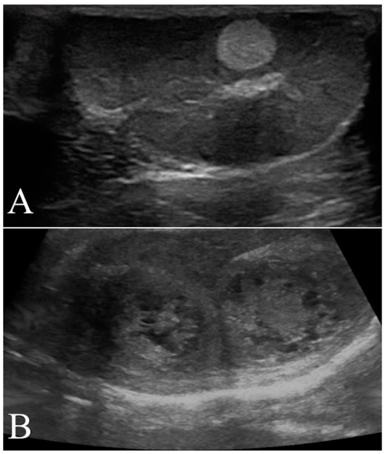
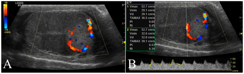
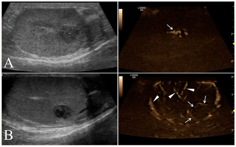
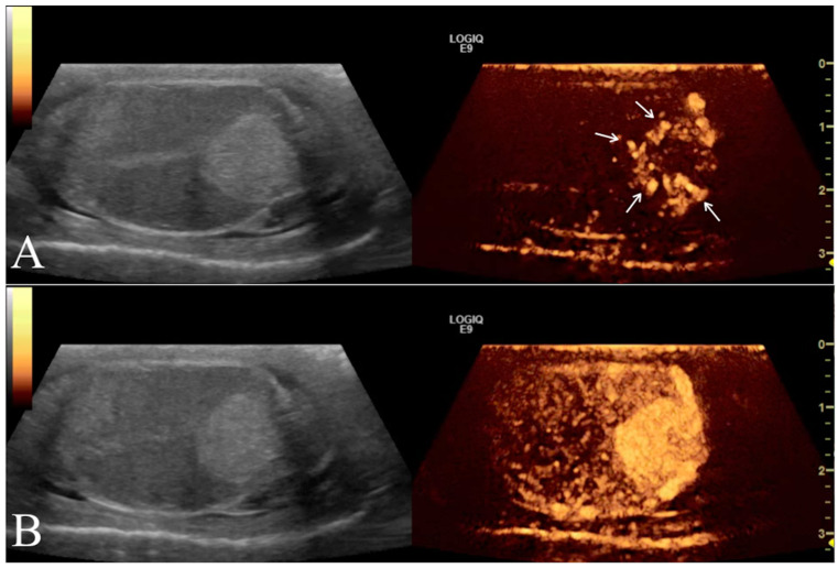
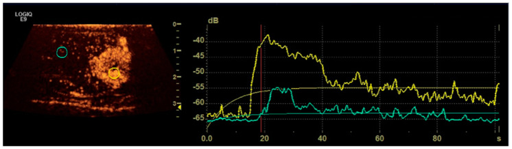
In this retrospective study, we assessed the accuracy of different blood flow imaging in diagnosing testicular tumor types in dogs. We recruited 27 dogs with leydigomas (14), seminomas (eight), sertoliomas (six), and mixed cells (five) confirmed histopathologically. In intact dogs, Pampiniform plexus and marginal arteries were scanned through pulsed Doppler. Blood flow and presence of intralesional/perilesional arteries were assessed by color and power Doppler, B-flow, and contrast-enhanced ultrasound. Tumor types did not differ by B-Mode ultrasonography characters. Pampiniform and testicular arteries of sertoliomas had higher (p < 0.05) pulsatility and resistive indexes. The proportion of leydigomas with a perilesional and/or perilesional/intralesional blood flow pattern detected by color and pulsed Doppler and B-flow was higher (p < 0.05) than that of the other tumors counted together. This resulted in a sensitivity of 81.8%, 83.3%, and 85.7%, a specificity of 76.5%, 56.3%, and 73.7%, and a correct classification rate of 78.6%, 67.9%, and 78.8%, respectively. While contrast enhanced ultrasound was highly effective in detecting all tumors, qualitative and quantitative parameters did not contribute to their differential diagnosis. In conclusion, results indicate that different testicular tumor types of dogs have subtly different vascular patterns, a condition that could help in identifying leydigomas.

Keywords: B-flow; CEUS; canine tumors; color Doppler; testes; ultrasonography.


Conflict of interest statement

The authors declare no conflict of interest.

Figures

Figure 1
Figure 1
Representative B-Mode ultrasonography scans. (A) Left testis of a 11 year old mixed breed dog with a focal hyperechoic nodule (seminoma) characterized by homogenous echotexture and regular margin. (B) Mixed cell tumor (leydig and seminoma) recorded as two nodular lesions in the right testis of a 13 year old Epagneul Breton. The lesions had mixed patterns, inhomogeneous echotextures and irregular margins.
Figure 2
Figure 2
Representative Doppler characterization of a nodular lesion (leydigoma) found in the left testicle of a 11 year old Italian Mastiff. In particular, picture 2 (A), recorded by color Doppler, allowed the visualization of blood flow distributed peripherally to the lesion, while picture 2 (B) showed the same blood flow sampled by pulsed wave mode for quantitative analysis.
Figure 3
Figure 3
Conventional ultrasonographic scans (left panels) of two different seminomas found respectively in the right testicle of a 10 year old Labrador Retriever in (A) and in the right testicle of a 9.5 year old German shepherd in (B). In the right panel the B-flow images show the I distribution of the lesional vessels (arrow) scored as 2 in (A), while in (B) it was possible to record the P pattern of vessels distribution (arrows) scored in this case as 1. In this B-flow image, it is also possible to visualize some physiological testicular vessels (arrows head), represented by the marginal region and intra-testicular branches of testicular artery.
Figure 4
Figure 4
Left testicle leydigoma of a 11 year old Italian Mastiff (the same of Figure 2). The left panel shows the sagittal B-mode ultrasound highlighting a focal hyperechoic nodule with homogeneous echotexture and regular margin. Representative contrast-enhanced ultrasound images of different contrast distribution phases are represented in the right panels. After 16 s from contrast injection it is possible to appreciate an early distribution of the contrast within the lesions (Arrows) compared to the surrounding parenchyma (A). Later (25 s) the contrast medium distributes homogeneously within the lesion that remain hyperenhanced compared to the normal testicle (B).
Figure 5
Figure 5
Representative image showing quantitative CEUS examination with ROIs positioned within the lesion (yellow ring) and in the normal parenchima (blue ring) respectively on the left and corresponding time-intensity curves in the right.

References

    1. Reifinger M. Statistical studies of the occurrence of testicular neoplasms in domestic mammals [Statistische Untersuchungen zum Vorkommen von Hodentumoren bei aussaugetieren] Zent. Vet. Reihe A. 1988;35:63–72. doi: 10.1111/j.1439-0442.1988.tb00007.x. - DOI - PubMed
    1. Mosier J.E. Effect of aging on body systems of the dog. Vet. Clin. N. Am. Small Anim. Pract. 1989;19:1–12. doi: 10.1016/S0195-5616(89)50001-9. - DOI - PubMed
    1. Lawrence J.A., Saba C.F. Tumors of the Male Reproductive System. In: Withrow S., Vail D., Page R., editors. Withrow & MacEwen’s Small Animal Clinical Oncology. 5th ed. Volume 28. Elsevier Saunders; Philadelphia, PA, USA: 2013. pp. 557–571.
    1. Grieco V., Riccardi E., Greppi G.F., Teruzzi F., Iermanò V., Finazzi M. Canine testicular tumours: A study on 232 dogs. J. Comp. Pathol. 2008;138:86–89. doi: 10.1016/j.jcpa.2007.11.002. - DOI - PubMed
    1. Grantham J.G., Charboneau J.W., James E.M., Kirschling R.J., Kvols L.K., Segura J.W., Wold L.E. Testicular neoplasms: 29 tumors studied by high resolution US. Radiology. 1985;157:775–780. doi: 10.1148/radiology.157.3.2997838. - DOI - PubMed

LinkOut - more resources