Review
. 2022 Jan 11;9(1):5.
doi: 10.3390/medicines9010005.

Diaphragm Ultrasound in Cardiac Surgery: State of the Art

Affiliations
Review

Diaphragm Ultrasound in Cardiac Surgery: State of the Art

Abdallah Fayssoil et al. Medicines (Basel). .

Abstract

In cardiac surgery, patients are at risk of phrenic nerve injury, which leads to diaphragm dysfunction and acute respiratory failure. Diaphragm dysfunction (DD) is relatively frequent in cardiac surgery and particularly affects patients after coronary artery bypass graft. The onset of DD affects patients' prognosis in term of weaning from mechanical ventilation and hospital length of stay. The authors present a narrative review about diaphragm physiology, techniques used to assess diaphragm function, and the clinical application of diaphragm ultrasound in patients undergoing cardiac surgery.

Keywords: cardiac ICU; cardiac surgery; diaphragm ultrasound; phrenic nerve.


Conflict of interest statement

The authors declare no conflict of interest.

Figures

Figure 1
Figure 1
Right diaphragm ultrasound. Note the normal inspiratory motion (64 mm) of the hemidiaphragm from the subcostal view. After the visualization of the right hemidiaphragm (bright line) using a B-mode (image on the left), an M mode was applied (image on the right) to record diaphragm motion.
Figure 2
Figure 2
Measurement of the right peak sniff tissue Doppler imaging velocity from the subcostal view. The diaphragm velocity was recorded during a sniff maneuver. Here is a reduced peak sniff velocity (7 cm/s) in a patient with muscular dystrophy.
Figure 3
Figure 3
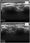
Measurement of the right diaphragm thickness (dotted line) in the end-expiratory phase (A) and end-inspiratory phase (B), using ultrasound. The diaphragm is visualized as a hypo-echogenic central layer surrounded by two hyper-echogenic lines, namely, the pleural line and the peritoneum.
Figure 4
Figure 4
Chest X ray in a post-operative patient with diaphragm paralysis. Note ascension of the left diaphragm.

References

    1. Bruni A., Garofalo E., Pasin L., Serraino G.F., Cammarota G., Longhini F., Landoni G., Lembo R., Mastroroberto P., Navalesi P., et al. Diaphragmatic Dysfunction after Elective Cardiac Surgery: A Prospective Observational Study. J. Cardiothorac. Vasc. Anesth. 2020;34:3336–3344. doi: 10.1053/j.jvca.2020.06.038. - DOI - PubMed
    1. Cavayas Y.A., Eljaiek R., Rodrigue É., Lamarche Y., Girard M., Wang H.T., Levesque S., Denault A.Y. Preoperative Diaphragm Function is Associated with Postoperative Pulmonary Complications after Cardiac Surgery. Crit. Care Med. 2019;47:e966–e974. doi: 10.1097/CCM.0000000000004027. - DOI - PubMed
    1. Diehl J.L., Lofaso F., Deleuze P., Similowski T., Lemaire F., Brochard L. Clinically relevant diaphragmatic dysfunction after cardiac operations. J. Thorac. Cardiovasc. Surg. 1994;107:487–498. doi: 10.1016/S0022-5223(94)70094-X. - DOI - PubMed
    1. Laghlam D., Lê M.P., Srour A., Monsonego R., Estagnasié P., Brusset A., Squara P. Diaphragm Dysfunction After Cardiac Surgery: Reappraisal. J. Cardiothorac. Vasc. Anesth. 2021;35:3241–3247. doi: 10.1053/j.jvca.2021.02.023. - DOI - PubMed
    1. Winkelmann E.R., Steffens É., Windmöller P., Fontela P.C., da Cruz D.T., Battisti I.D.E. Preoperative expiratory and inspiratory muscle weakness to predict postoperative outcomes in patients undergoing elective cardiac surgery. J. Card. Surg. 2020;35:128–134. doi: 10.1111/jocs.14355. - DOI - PubMed

LinkOut - more resources