

Hao1 Is Not a Pathogenic Factor for Ectopic Ossifications but Functions to Regulate the TCA Cycle In Vivo
- PMID: 35050204
- PMCID: PMC8780519
- DOI: 10.3390/metabo12010082
Hao1 Is Not a Pathogenic Factor for Ectopic Ossifications but Functions to Regulate the TCA Cycle In Vivo
Abstract
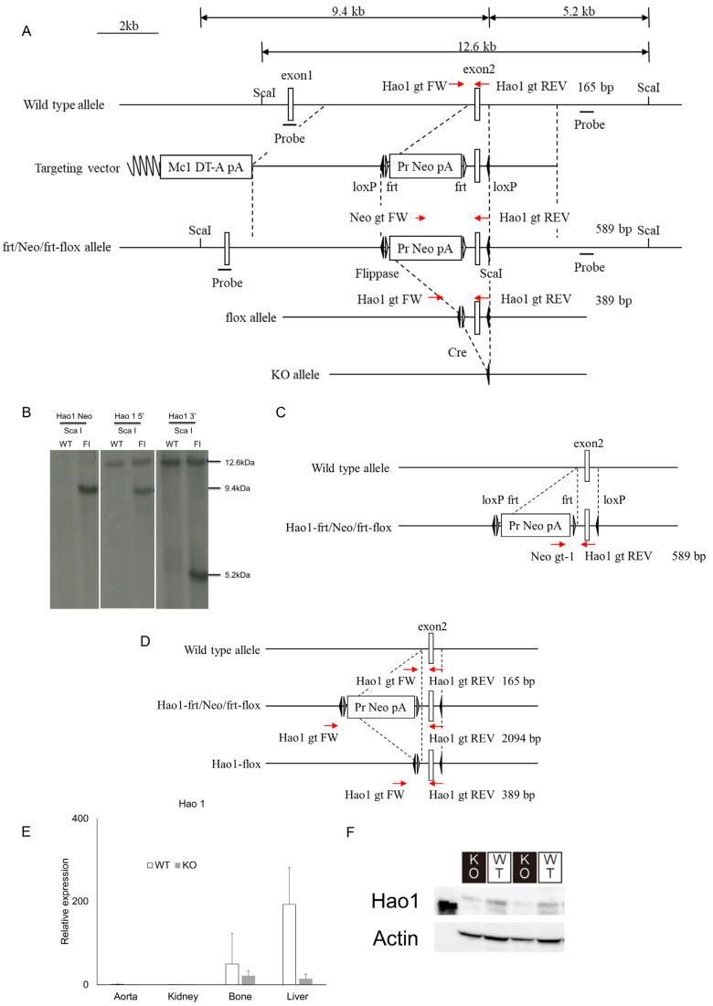
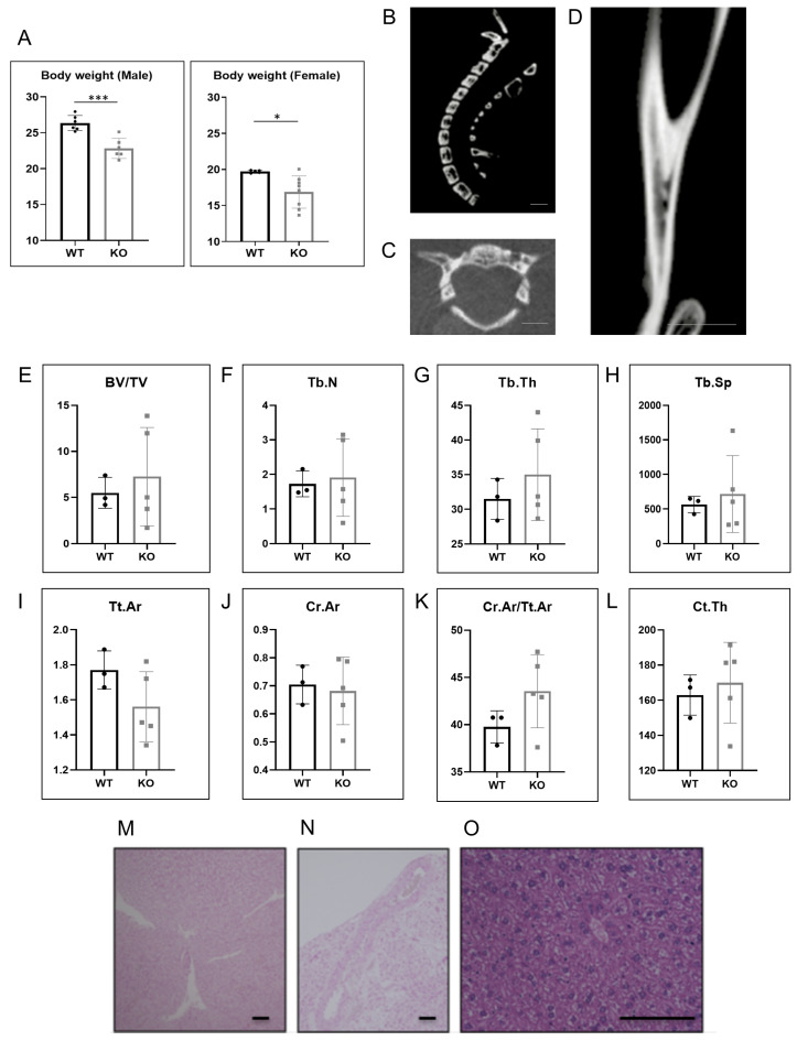
Ossification of the posterior longitudinal ligament (OPLL), a disease characterized by the ectopic ossification of a spinal ligament, promotes neurological disorders associated with spinal canal stenosis. While blocking ectopic ossification is mandatory to prevent OPLL development and progression, the mechanisms underlying the condition remain unknown. Here we show that expression of hydroxyacid oxidase 1 (Hao1), a gene identified in a previous genome-wide association study (GWAS) as an OPLL-associated candidate gene, specifically and significantly decreased in fibroblasts during osteoblast differentiation. We then newly established Hao1-deficient mice by generating Hao1-flox mice and crossing them with CAG-Cre mice to yield global Hao1-knockout (CAG-Cre/Hao1flox/flox; Hao1 KO) animals. Hao1 KO mice were born normally and exhibited no obvious phenotypes, including growth retardation. Moreover, Hao1 KO mice did not exhibit ectopic ossification or calcification. However, urinary levels of some metabolites of the tricarboxylic acid (TCA) cycle were significantly lower in Hao1 KO compared to control mice based on comprehensive metabolomic analysis. Our data indicate that Hao1 loss does not promote ectopic ossification, but rather that Hao1 functions to regulate the TCA cycle in vivo.
Keywords: ectopic ossification; hydroxyacid oxidase 1; ossification of the posterior longitudinal ligament; tricarboxylic acid cycle.
Conflict of interest statement
All authors state that they have no conflicts of interest with the contents of this article.
Figures


Similar articles
-
Expression Analysis of Susceptibility Genes for Ossification of the Posterior Longitudinal Ligament of the Cervical Spine in Human OPLL-related Tissues and a Spinal Hyperostotic Mouse (ttw/ttw).Spine (Phila Pa 1976). 2020 Nov 15;45(22):E1460-E1468. doi: 10.1097/BRS.0000000000003648. Spine (Phila Pa 1976). 2020. PMID: 32756283
-
Runx2 haploinsufficiency ameliorates the development of ossification of the posterior longitudinal ligament.PLoS One. 2012;7(8):e43372. doi: 10.1371/journal.pone.0043372. Epub 2012 Aug 21. PLoS One. 2012. PMID: 22927960 Free PMC article.
-
Upregulated expression of connexin43 in spinal ligament fibroblasts derived from patients presenting ossification of the posterior longitudinal ligament.Spine (Phila Pa 1976). 2011 Dec 15;36(26):2267-74. doi: 10.1097/BRS.0b013e31820ccfc6. Spine (Phila Pa 1976). 2011. PMID: 21311398
-
Pharmacological aspect of ectopic ossification in spinal ligament tissues.Pharmacol Ther. 2008 Jun;118(3):352-8. doi: 10.1016/j.pharmthera.2008.03.007. Epub 2008 Apr 6. Pharmacol Ther. 2008. PMID: 18499263 Review.
-
Comparison of clinical outcomes in decompression and fusion versus decompression only in patients with ossification of the posterior longitudinal ligament: a meta-analysis.Neurosurg Focus. 2016 Jun;40(6):E9. doi: 10.3171/2016.3.FOCUS1630. Neurosurg Focus. 2016. PMID: 27246492
Cited by
-
Analysis of the Protective Effects of Rosa roxburghii-Fermented Juice on Lipopolysaccharide-Induced Acute Lung Injury in Mice through Network Pharmacology and Metabolomics.Nutrients. 2024 Apr 30;16(9):1376. doi: 10.3390/nu16091376. Nutrients. 2024. PMID: 38732622 Free PMC article.
-
Integration of whole genome resequencing and transcriptome sequencing to identify candidate genes for tall and short traits in Baicheng Fatty chickens.Front Vet Sci. 2025 Feb 27;12:1534742. doi: 10.3389/fvets.2025.1534742. eCollection 2025. Front Vet Sci. 2025. PMID: 40084164 Free PMC article.
-
Establishment and validation of a novel peroxisome-related gene prognostic risk model in kidney clear cell carcinoma.BMC Urol. 2024 Jan 31;24(1):26. doi: 10.1186/s12894-024-01404-z. BMC Urol. 2024. PMID: 38297313 Free PMC article.
-
Screening of potential biomarkers in propofol-induced neurotoxicity via bioinformatics prediction and experimental verification.Am J Transl Res. 2024 Mar 15;16(3):755-767. doi: 10.62347/MTAY7931. eCollection 2024. Am J Transl Res. 2024. PMID: 38586100 Free PMC article.
References
-
- Kawaguchi Y., Nakano M., Yasuda T., Seki S., Hori T., Suzuki K., Makino H., Kimura T. Characteristics of ossification of the spinal ligament; incidence of ossification of the ligamentum flavum in patients with cervical ossification of the posterior longitudinal ligament—Analysis of the whole spine using multidetector CT. J. Orthop. Sci. 2016;21:439–445. doi: 10.1016/j.jos.2016.04.009. - DOI - PubMed
-
- Koyanagi I., Iwasaki Y., Hida K., Imamura H., Fujimoto S., Akino M. Acute cervical cord injury associated with ossification of the posterior longitudinal ligament. Neurosurgery. 2003;53:887–891; discussion 891–882. doi: 10.1227/01.NEU.0000083590.84053.CC. - DOI - PubMed
LinkOut - more resources
Full Text Sources
Research Materials
