. 2021 Dec 21;9(1):1.
doi: 10.3390/jcdd9010001.

Characterizing Neonatal Heart Maturation, Regeneration, and Scar Resolution Using Spatial Transcriptomics

Affiliations

Characterizing Neonatal Heart Maturation, Regeneration, and Scar Resolution Using Spatial Transcriptomics

Adwiteeya Misra et al. J Cardiovasc Dev Dis. .

Abstract

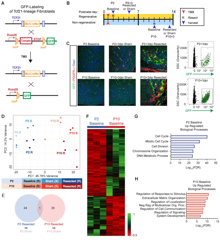
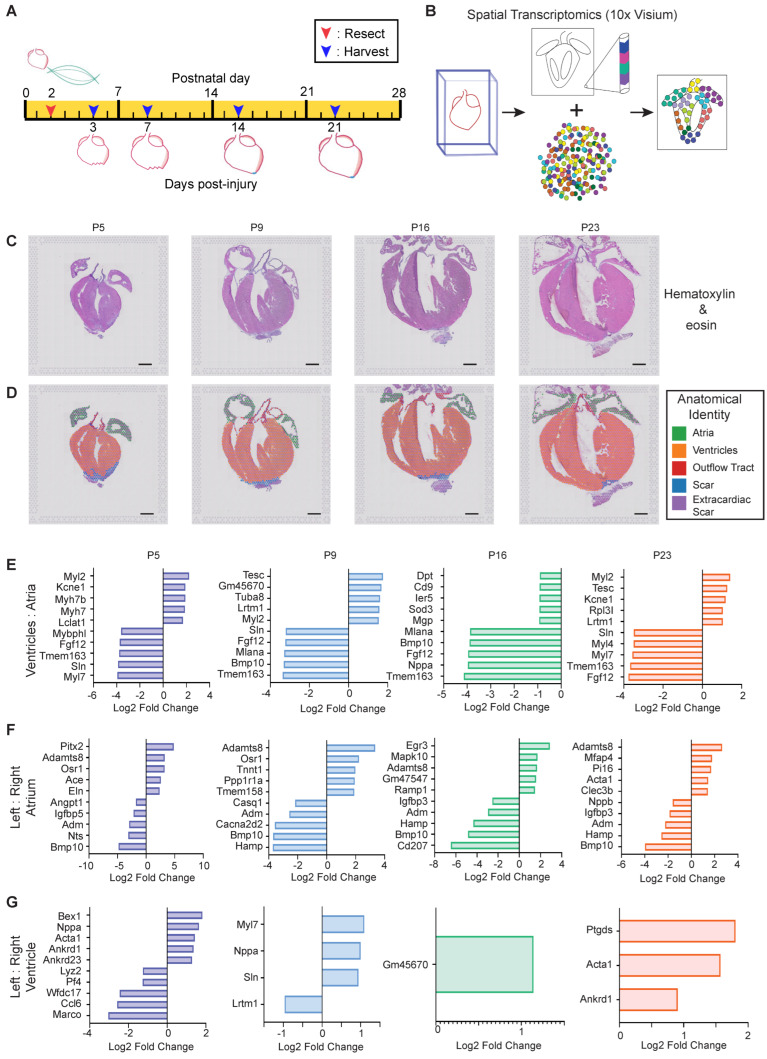
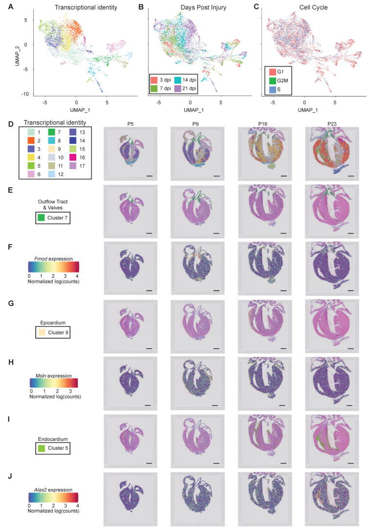
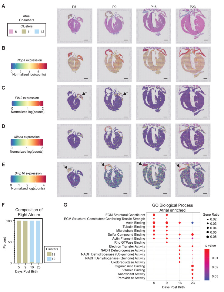
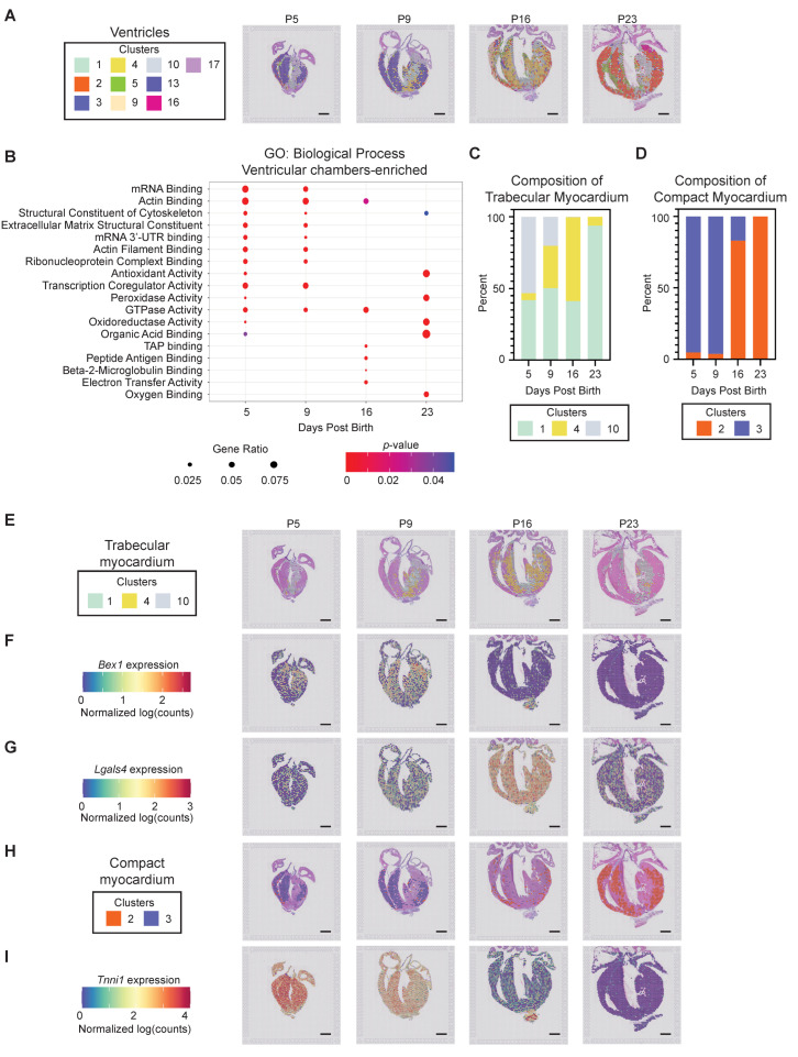
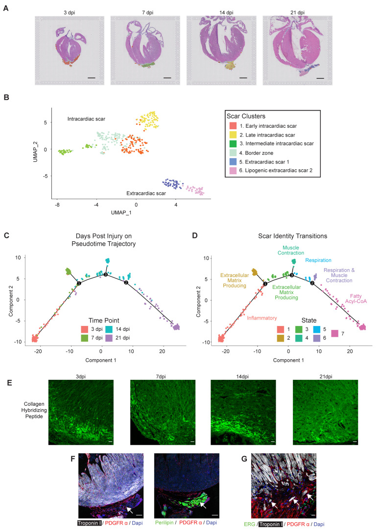
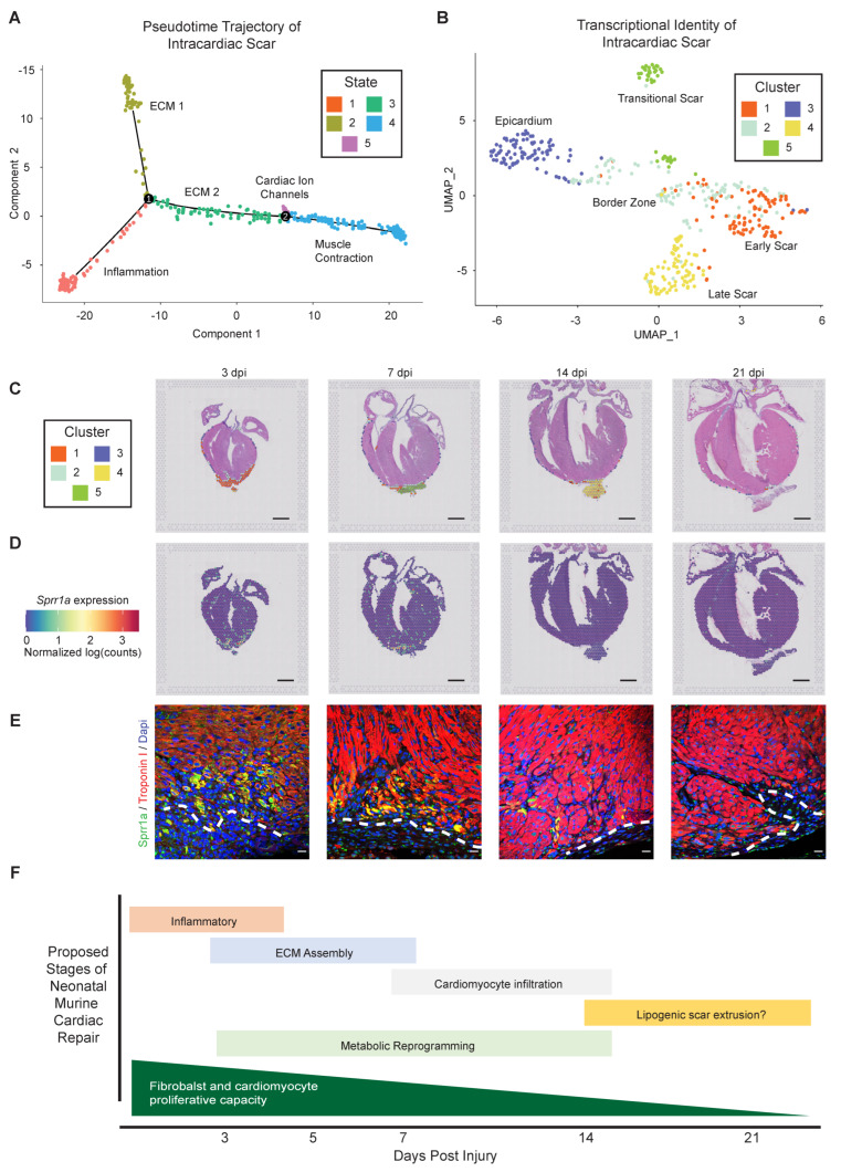
The neonatal mammalian heart exhibits a remarkable regenerative potential, which includes fibrotic scar resolution and the generation of new cardiomyocytes. To investigate the mechanisms facilitating heart repair after apical resection in neonatal mice, we conducted bulk and spatial transcriptomic analyses at regenerative and non-regenerative timepoints. Importantly, spatial transcriptomics provided near single-cell resolution, revealing distinct domains of atrial and ventricular myocardium that exhibit dynamic phenotypic alterations during postnatal heart maturation. Spatial transcriptomics also defined the cardiac scar, which transitions from a proliferative to secretory phenotype as the heart loses regenerative potential. The resolving scar is characterized by spatially and temporally restricted programs of inflammation, epicardium expansion and extracellular matrix production, metabolic reprogramming, lipogenic scar extrusion, and cardiomyocyte restoration. Finally, this study revealed the emergence of a regenerative border zone defined by immature cardiomyocyte markers and the robust expression of Sprr1a. Taken together, our study defines the spatially and temporally restricted gene programs that underlie neonatal heart regeneration and provides insight into cardio-restorative mechanisms supporting scar resolution.

Keywords: fibroblast; heart; mouse; regeneration; scar; spatial transcriptomics.


Conflict of interest statement

The authors declare no conflict of interest.

Figures

Figure 1
Figure 1
RNA sequencing of resident cardiac fibroblasts reveals a switch to secretory phenotype which corresponds to loss of regenerative capacity. (A) Schematic describing the breeding strategy used to generate mice that allowed fluorescent lineage training of resident fibroblasts. (B) Schematic describing experimental strategy, including timing of tamoxifen (TMX) injections, apical resection or sham surgeries (R), and fibroblast and tissue isolations. (C) Hearts were obtained from postnatal mice at indicated timepoints, and stained with antibodies to visualize GFP (Tcf21+ fibroblast lineage), PDGFR-a (endothelial cells and fibroblasts) and DAPI (nuclei). Representative flow cytometry plots are shown on the right that indicate the population of GFP+ fibroblasts obtained for RNA-sequencing. (D) Principal component analysis (PCA), revealing independent gene expression programs based on RNA-sequencing of cardiac fibroblasts of indicated treatment. (E) Venn diagram representation of differentially expressed genes in response to apical resection at indicated timepoints. (F) Heat map representation of genes that exhibit differential expression between P2 and P10 at baseline. (G) Biological processes that are enriched in cardiac fibroblasts at P2, compared to P10. (H) Biological processes that are enriched in cardiac fibroblasts at P10 compared to P2.
Figure 2
Figure 2
Spatially and temporally resolved transcriptional map of the regenerating neonatal mouse heart. (A) Schematic representation of the experimental timeline of apical resection (R) and tissue harvest for spatial transcriptomics. (B) 10X Visium spatial transcriptomics experimental strategy. A single representative heart was evaluated by spatial transcriptomics for each timepoint on a single slide. (C) Hematoxylin & Eosin stained sections used for spatial transcriptomics. (D) Loupe Browser was used to assign regional posts an anatomical identity, indicated by color. (EG) Rank ordering of differentially expressed genes at timepoints and comparison indicated in the graph reveals the most-enriched genes that define the (E) ventricles versus atria; (F) left versus right atrium; or (G) left versus right ventricle. Scale bar = 1 mm.
Figure 3
Figure 3
Spatial transcriptomics reveals spatially restricted gene programs in postnatal mouse heart regeneration at near single-cell resolution. (A) Uniform manifold approximation and projection (UMAP) was generated using Seurat to visualize barcoded spots with similar transcriptional identity based on RNA-sequencing. (B,C) Barcoded spots within UMAP were labeled based on (B) days postinjury (dpi); and (C) phase of the cell cycle. (D) Barcoded spots representing 17 transcriptionally distinct clusters were mapped to their origin within a tissue section for the unbiased identification of anatomical structures with unique gene expression programs. (E) Localization of cluster 7 spots reveals localization to the outflow tract and valves. (F) Spatially resolved Fmod expression reveals enrichment in heart valves. (G) Localization of cluster 9 spots reveals localization to the epicardium. Note the single layer of spots that labels the epicardium on the heart’s surface, and the epicardial expansion observed at 14 dpi. (H) Spatially resolved Msln expression reveals enrichment in heart valves. (I) Localization of cluster 5 spots reveals localization to the endocardium. (J) Spatially resolved Alas2 expression reveals enrichment in endocardium, especially as heart maturation proceeds. Scale bar = 1mm.
Figure 4
Figure 4
Spatial transcriptomics defines postnatal program of atrial chamber maturation. (A) Barcoded spots representing the 3 transcriptionally distinct atrial clusters were mapped to their origin. (B–E) Spatially resolved expression of atrial enriched genes identified by spatial transcriptomics reveals (B) Nppa enrichment in both atria at all timepoints, and trabecular myocardium at P5 and P9; (C) Pitx2 enrichment in the left atrium, especially at P5 and P9; (D) Mlana expression enriched in the right atria, especially after P5; and (E) Bmp10 enrichment in the right atrium at all timepoints. Arrows indicate left (C) or right (E) atrium. (F) Right atrium maturation program is revealed consisting of a shift from cluster 11 identity at P5 and P9 to cluster 12 identity at P16 and P23. (G) Biological processes were interrogated as a function of postnatal time, revealing a shift from ECM and actin/microtubule processes to oxidative metabolism as the atria mature. Scale bar = 1 mm.
Figure 5
Figure 5
Spatially resolved transcriptional programs define trabecular and compact ventricular myocardium maturation. (A) Barcoded spots, representing the 10 transcriptionally distinct ventricular clusters, were mapped to their origin. (B) Biological processes were interrogated as a function of postnatal time, revealing a shift from ECM, actin binding, and cytoskeleton processes to oxidative metabolism as the ventricles mature. (C) Trabecular myocardium maturation program is revealed, consisting of a shift from 1 and 10 at P5, through an intermediate identity with a high percentage of cluster 4, to being dominated by cluster 1 at P23. (D) Compact myocardium maturation program is revealed, consisting of a shift from cluster 3 identity at P5 and P9 to cluster 2 identity at P16 and P23. (E) Barcoded spots with trabecular myocardium identity were mapped back to an anatomical origin. (F,G) Spatially resolved expression of trabecular enriched genes identified by spatial transcriptomics reveals (F) Bex1 expression within trabecular myocardium, especially at P5 and P9; and (G) Lgals4 expression, particularly at the intermediate P16 timepoint. (H) Barcoded spots with compact myocardium identity were mapped back to an anatomical origin. (I) Spatially resolved Tnni1 expression reveals a decrease within compact and trabecular myocardium as the heart matures. Scale bar = 1 mm.
Figure 6
Figure 6
Spatial transcriptomics reveals the phenotypic trajectory of the regenerative scar, consisting of bi-phasic collagen deposition and culminating in scar extrusion. (A) Barcoded spots with scar identity were mapped back to an anatomical origin. (B) Uniform manifold approximation and projection (UMAP) was generated using Seurat to visualize 6 regional identities within the regenerative scar. (C) The developmental trajectory of scar-associated spots was integrated with days post-injury (dpi). (D) Pseudotime trajectory was integrated with GO term enrichments to establish phenotypic timeline. (E) Representative heart sections obtained at the indicated time after apical resection surgery were stained with collagen hybridizing peptide (CHP) to visualize nascent and remodeling collagen at the cardiac apex. This trend was observed in n = 3/5 independent samples (3 dpi) and n = 4/4 independent samples (14 dpi). (F,G) Representative heart sections obtained at 21 dpi were stained with antibodies directed against Troponin I (cardiomyocytes, white), PDGFR-a (fibroblasts, red) and perilipin (adipocyte, green in left image) and ERG (endothelial cells, green right image). DAPI stains nuclei (blue). Note the extracardiac fibro-lipogenic scar emerging from the apex (left), that often harbors individual cardiomyocytes (white arrows, right image). Scale bar = 1 mm (A) and 100 mm (EG).
Figure 7
Figure 7
Spatial transcriptomics reveals the phenotypic trajectory of the intracardiac resolving intracardiac scar and border zone. (A) Pseudotime developmental trajectory was integrated with biological process enrichment to identify the regional identity of the 5 intracardiac scar states. (B) Uniform manifold approximation and projection reveals 5 distinct transcriptional clusters that define the temporal progression of the intracardiac scar, the border zone, and the epicardium. (C) Barcoded spots were mapped back to an anatomical origin to spatially and temporally resolve the intracardiac scar identity, and location of the border zone as a narrow band of tissue between the injury and healthy myocardium. (D) Spatially resolved Sprr1a expression reveals an enrichment within the border zone specifically at 3 and 7 dpi. (E) Representative heart sections obtained at indicated post-injury timepoints were stained with antibodies directed against Troponin I (cardiomyocytes, red) and Sprr1a (green). DAPI labels nuclei, and the dashed line indicates transition from injury to healthy myocardium, defined as the border zone. Note the enrichment of Sprr1a in border zone cardiomyocytes at 3 dpi and 7 dpi, which is absent in remote myocardium. (F) Schematic of the proposed stages of neonatal mouse heart regeneration, based on bulk RNA-sequencing of isolated fibroblasts and spatial transcriptomics at key regenerative timepoints. Scale bar = 1 mm (C,D) and 100 mm (E).

References

    1. Frangogiannis N.G. The Extracellular Matrix in Ischemic and Nonischemic Heart Failure. Circ. Res. 2019;125:117–146. doi: 10.1161/CIRCRESAHA.119.311148. - DOI - PMC - PubMed
    1. Virani S.S., Alonso A., Aparicio H.J., Benjamin E.J., Bittencourt M.S., Callaway C.W., Carson A.P., Chamberlain A.M., Cheng S., Delling F.N., et al. Heart Disease and Stroke Statistics-2021 Update: A Report From the American Heart Association. Circulation. 2021;143:e254–e743. doi: 10.1161/CIR.0000000000000950. - DOI - PubMed
    1. Mahmoud A.I., Porrello E., Kimura W., Olson E.N., Sadek H.A. Surgical models for cardiac regeneration in neonatal mice. Nat. Protoc. 2014;9:305–311. doi: 10.1038/nprot.2014.021. - DOI - PMC - PubMed
    1. Polizzotti B.D., Ganapathy B., Haubner B.J., Penninger J., Kühn B.D.P.B.G.B. A cryoinjury model in neonatal mice for cardiac translational and regeneration research. Nat. Protoc. 2016;11:542–552. doi: 10.1038/nprot.2016.031. - DOI - PMC - PubMed
    1. Porrello E.R., Mahmoud A.I., Simpson E., Hill J.A., Richardson J.A., Olson E.N., Sadek H. Transient Regenerative Potential of the Neonatal Mouse Heart. Science. 2011;331:1078–1080. doi: 10.1126/science.1200708. - DOI - PMC - PubMed