. 2022 Jan 3;63(1):23.
doi: 10.1167/iovs.63.1.23.

Autofluorescent Organelles Within the Retinal Pigment Epithelium in Human Donor Eyes With and Without Age-Related Macular Degeneration

Affiliations

Autofluorescent Organelles Within the Retinal Pigment Epithelium in Human Donor Eyes With and Without Age-Related Macular Degeneration

Katharina Bermond et al. Invest Ophthalmol Vis Sci. .

Abstract

Purpose: Human retinal pigment epithelium (RPE) cells contain lipofuscin, melanolipofuscin, and melanosome organelles that impact clinical autofluorescence (AF) imaging. Here, we quantified the effect of age-related macular degeneration (AMD) on granule count and histologic AF of RPE cell bodies.

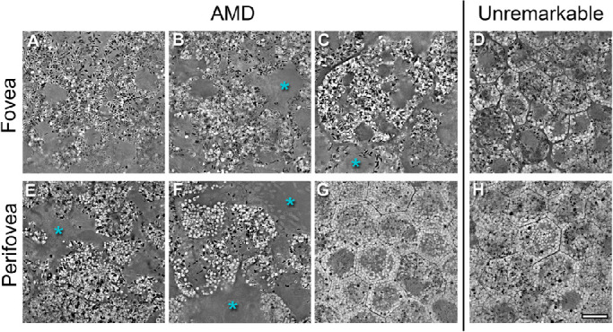
Methods: Seven AMD-affected human RPE-Bruch's membrane flatmounts (early and intermediate = 3, late dry = 1, and neovascular = 3) were imaged at fovea, perifovea, and near periphery using structured illumination and confocal AF microscopy (excitation 488 nm) and compared to RPE-flatmounts with unremarkable macula (n = 7, >80 years). Subsequently, granules were marked with computer assistance, and classified by their AF properties. The AF/cell was calculated from confocal images. The total number of granules and AF/cell was analyzed implementing a mixed effect analysis of covariance (ANCOVA).

Results: A total of 152 AMD-affected RPE cells were analyzed (fovea = 22, perifovea = 60, and near-periphery = 70). AMD-affected RPE cells showed increased variability in size and a significantly increased granule load independent of the retinal location (fovea: P = 0.02, perifovea: P = 0.04, and near periphery: P < 0.01). The lipofuscin fraction of total organelles decreased and the melanolipofuscin fraction increased in AMD, at all locations (especially the fovea). AF was significantly lower in AMD-affected cells (fovea: <0.01, perifovea: <0.01, and near periphery: 0.02).

Conclusions: In AMD RPE, lipofuscin was proportionately lowest in the fovea, a location also known to be affected by accumulation of soft drusen and preservation of cone-mediated visual acuity. Enlarged RPE cell bodies displayed increased net granule count but diminished total AF. Future studies should also assess the impact on AF imaging of RPE apical processes containing melanosomes.


Conflict of interest statement

Disclosure: K. Bermond, None; L. von der Emde, None; I.-S. Tarau, None; L. Bourauel, None; R. Heintzmann, None; F.G. Holz, Acucela (C, F), Allergan (F), Apellis (C, F), Bayer (C, F), Boehringer-Ingelheim (C), Bioeq/Formycon (F, C), CenterVue (F), Ellex (F), Roche/Genentech (C, F), Geuder (C, F), Graybug (C), Gyroscope (C), Heidelberg Engineering (C, F), IvericBio (C, F), Kanghong (C, F), LinBioscience (C), NightStarX (F), Novartis (C, F), Optos (F), Oxurion (C), Pixium Vision (C, F), Oxurion (C), Stealth BioTherapeutics (C), Zeiss (F, C); C.A. Curcio, Genentech (F), Regeneron (F), MacRegen Inc. (I); K.R. Sloan, MacRegen Inc. (I); T. Ach, Roche (C), Novartis (C), Novartis (R), MacRegen Inc. (I)

Figures

Figure 1.
Figure 1.
SIM-images of AMD-affected and unremarkable tissue. Each image shows a representative slide at the apical surface of a SIM z-stack of the fovea (A–D) or perifovea (E–H) of AMD-affected (A–C, E–G) or unremarkable (D, H) tissues. The AMD-affected tissues show deterioration of cell structures like dissolving cell borders, partial loss of intracellular granule organization and cell loss. Areas of RPE cell loss probably associated with deposits (basal laminar deposits/basal linear deposits) are marked with a blue asterisk. These deposits affected the fovea and, in some tissues, extended to the perifovea (E, F). Where the perifovea was not (yet) affected by AMD (G), the tissue appeared comparable to healthy tissue (D, H) which shows uniform cell shapes and orderly intracellular granule distribution. Note: the granule free areas within the cell bodies in D and H represent the non-autofluorescent cell nucleus. In panels A, B, and E, we were unable to distinguish cell boundaries and used cell-size equivalent squares instead. A 3D-version of panel C is further depicted in the supplemental video. Early or intermediate AMD: 83-year-old woman (F), 84-year-old man B, and 86-year-old woman A. Late non-exudative AMD: 81-year-old man C, G. Late exudative AMD: 90-year-old woman E. Unremarkable tissue: 83-year-old woman D and 90-year-old woman H. Scale bar = 10 µm.
Figure 2.
Figure 2.
En face (A) and cross section (B) view of intracellular granule distribution. Each analyzed granule was color-coded and plotted (lipofuscin: yellow, melanolipofuscin 1–3: blue, melanolipofuscin 4: pink, melanolipofuscin 5: magenta, and M: orange). Only few melanosomes, melanolipofuscin 4, and melanolipofuscin 5 were detectable. Red lines represent cell borders or artificial squares, respectively. At four locations (three foveae and one perifovea), no granule analysis was possible due to large RPE-atrophy. Foveal tissue showed distinctive alterations in cell structure with either formation of enlarged cells (83-year-old woman and 81-year-old man) or primarily faded cell borders and a moth-eaten appearance (84-year-old man and 86-year-old woman [early/intermediate AMD]). The predominant granule type at the fovea remained melanolipofuscin, like in unremarkable tissues. At the perifovea and near periphery, all tissues that showed a regular cell structure in the en face view also displayed two bands in the cross-section view: an apical band with predominantly melanolipofuscin and lipofuscin and a basal band with predominantly lipofuscin. At locations where the cell structures were deteriorated (83-year-old perifovea and near periphery, 90-year-old perifovea) these two bands could not be distinguished. Scale bar = 10 µm.
Figure 3.
Figure 3.
Granule load per cell area. The graph shows the area (x-axis) and total granule load (y-axis) of RPE cells from donors with age-related macular degeneration (top row) and aged donors with unremarkable macula (bottom row). Whereas aged RPE cells displayed a clear trend between cell area and granule load, AMD RPE cells demonstrated a dissociation between the two. Using repeated measures correlation to adjust for the dependence of different cells from one tissue we found a strong correlation between cell area and granule load in AMD RPE cells as well (perifovea: r = 0.77 [95% confidence interval [0.61, 0.87], P value < 0.001/near periphery: r = 0.86 [95% confidence interval 0.78, 0.91] P value < 0.001). These results show that the relationship for granule load and cell area is stronger within-tissue than between-tissue. RPE cells in AMD presented an increased cell area and granule load. This trend was most pronounced at the fovea. The graph also illustrates an increased variability in cell area and granule load in AMD.

References

    1. Strauss O. The retinal pigment epithelium in visual function. Physiol Rev. 2005; 85(3): 845–881. - PubMed
    1. Pollreisz A, Messinger JD, Sloan KR, et al. .. Visualizing melanosomes, lipofuscin, and melanolipofuscin in human retinal pigment epithelium using serial block face scanning electron microscopy. Exp Eye Res. 2018; 166: 131–139. - PubMed
    1. Pollreisz A, Neschi M, Sloan KR, et al. .. Atlas of Human Retinal Pigment Epithelium Organelles Significant for Clinical Imaging. Invest Ophthalmol Vis Sci. 2020; 61(8): 13. - PMC - PubMed
    1. Bermond K, Wobbe C, Tarau I-S, et al. .. Autofluorescent Granules of the Human Retinal Pigment Epithelium: Phenotypes, Intracellular Distribution, and Age-Related Topography. Invest Ophthalmol Vis Sci. 2020; 61(5): 35. - PMC - PubMed
    1. Feeney-Burns L, Hilderbrand ES, Eldridge S.. Aging human RPE: morphometric analysis of macular, equatorial, and peripheral cells. Invest Ophthalmol Vis Sci. 1984; 25(2): 195–200. - PubMed

Publication types